4LQW

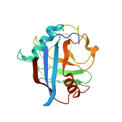
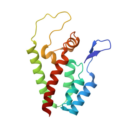
Crystal structure of HIV-1 capsid N-terminal domain in complex with NUP358 cyclophilin


Experimental Data Snapshot

  • Method: X-RAY DIFFRACTION
  • Resolution: 1.95 Å
  • R-Value Free: 0.246 
  • R-Value Work: 0.205 
  • R-Value Observed: 0.207 

Starting Model: experimental
View more details

wwPDB Validation   3D Report Full Report


This is version 1.2 of the entry. See complete history


Literature

HIV-1 capsid undergoes coupled binding and isomerization by the nuclear pore protein NUP358.

Bichel, K.Price, A.J.Schaller, T.Towers, G.J.Freund, S.M.James, L.C.

(2013) Retrovirology 10: 81-81

  • DOI: https://doi.org/10.1186/1742-4690-10-81
  • Primary Citation of Related Structures:  
    4LQW

  • PubMed Abstract: 

    Lentiviruses such as HIV-1 can be distinguished from other retroviruses by the cyclophilin A-binding loop in their capsid and their ability to infect non-dividing cells. Infection of non-dividing cells requires transport through the nuclear pore but how this is mediated is unknown. Here we present the crystal structure of the N-terminal capsid domain of HIV-1 in complex with the cyclophilin domain of nuclear pore protein NUP358. The structure reveals that HIV-1 is positioned to allow single-bond resonance stabilisation of exposed capsid residue P90. NMR exchange experiments demonstrate that NUP358 is an active isomerase, which efficiently catalyzes cis-trans isomerization of the HIV-1 capsid. In contrast, the distantly related feline lentivirus FIV can bind NUP358 but is neither isomerized by it nor requires it for infection. Isomerization by NUP358 may be preserved by HIV-1 to target the nuclear pore and synchronize nuclear entry with capsid uncoating.


  • Organizational Affiliation

    Protein and Nucleic Acid Chemistry Division, Medical Research Council Laboratory of Molecular Biology, Hills Road, Cambridge CB2 0QH, UK.


Macromolecules
Find similar proteins by:  (by identity cutoff)  |  3D Structure
Entity ID: 1
MoleculeChains Sequence LengthOrganismDetailsImage
E3 SUMO-protein ligase RanBP2
A, B
177Homo sapiensMutation(s): 0 
Gene Names: RANBP2NUP358
EC: 6.3.2 (PDB Primary Data), 5.2.1.8 (PDB Primary Data), 2.3.2 (UniProt)
UniProt & NIH Common Fund Data Resources
Find proteins for P49792 (Homo sapiens)
Explore P49792 
Go to UniProtKB:  P49792
PHAROS:  P49792
GTEx:  ENSG00000153201 
Entity Groups  
Sequence Clusters30% Identity50% Identity70% Identity90% Identity95% Identity100% Identity
UniProt GroupP49792
Sequence Annotations
Expand
  • Reference Sequence
Find similar proteins by:  (by identity cutoff)  |  3D Structure
Entity ID: 2
MoleculeChains Sequence LengthOrganismDetailsImage
Capsid protein p24
C, D
146Human immunodeficiency virus type 1 (NEW YORK-5 ISOLATE)Mutation(s): 0 
Gene Names: gag-pol
UniProt
Find proteins for P12497 (Human immunodeficiency virus type 1 group M subtype B (isolate NY5))
Explore P12497 
Go to UniProtKB:  P12497
Entity Groups  
Sequence Clusters30% Identity50% Identity70% Identity90% Identity95% Identity100% Identity
UniProt GroupP12497
Sequence Annotations
Expand
  • Reference Sequence
Experimental Data & Validation

Experimental Data

  • Method: X-RAY DIFFRACTION
  • Resolution: 1.95 Å
  • R-Value Free: 0.246 
  • R-Value Work: 0.205 
  • R-Value Observed: 0.207 
  • Space Group: C 1 2 1
Unit Cell:
Length ( Å )Angle ( ˚ )
a = 142.18α = 90
b = 38.26β = 100.64
c = 123.81γ = 90
Software Package:
Software NamePurpose
PHASERphasing
REFMACrefinement
MOSFLMdata reduction
SCALAdata scaling

Structure Validation

View Full Validation Report


Entry History 

Deposition Data

Revision History  (Full details and data files)

  • Version 1.0: 2013-08-14
    Type: Initial release
  • Version 1.1: 2015-02-04
    Changes: Structure summary
  • Version 1.2: 2023-09-20
    Changes: Data collection, Database references, Refinement description