4LPS | pdb_00004lps

Crystal structure of HypB from Helicobacter pylori in complex with nickel


Experimental Data Snapshot

  • Method: X-RAY DIFFRACTION
  • Resolution: 2.00 Å
  • R-Value Free: 
    0.196 (Depositor), 0.205 (DCC) 
  • R-Value Work: 
    0.170 (Depositor), 0.175 (DCC) 
  • R-Value Observed: 
    0.172 (Depositor) 

Starting Model: experimental
View more details

wwPDB Validation 3D Report Full Report

Validation slider image for 4LPS

Ligand Structure Quality Assessment 


This is version 1.2 of the entry. See complete history

Literature

Relationship between Ni(II) and Zn(II) Coordination and Nucleotide Binding by the Helicobacter pylori [NiFe]-Hydrogenase and Urease Maturation Factor HypB.

Sydor, A.M.Lebrette, H.Ariyakumaran, R.Cavazza, C.Zamble, D.B.

(2014) J Biological Chem 289: 3828-3841

  • DOI: https://doi.org/10.1074/jbc.M113.502781
  • Primary Citation Related Structures: 
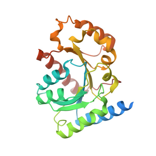
    4LPS

  • PubMed Abstract: 

    The pathogen Helicobacter pylori requires two nickel-containing enzymes, urease and [NiFe]-hydrogenase, for efficient colonization of the human gastric mucosa. These enzymes possess complex metallocenters that are assembled by teams of proteins in multistep pathways. One essential accessory protein is the GTPase HypB, which is required for Ni(II) delivery to [NiFe]-hydrogenase and participates in urease maturation. Ni(II) or Zn(II) binding to a site embedded in the GTPase domain of HypB modulates the enzymatic activity, suggesting a mechanism of regulation. In this study, biochemical and structural analyses of H. pylori HypB (HpHypB) revealed an intricate link between nucleotide and metal binding. HpHypB nickel coordination, stoichiometry, and affinity were modulated by GTP and GDP, an effect not observed for zinc, and biochemical evidence suggests that His-107 coordination to nickel toggles on and off in a nucleotide-dependent manner. These results are consistent with the crystal structure of HpHypB loaded with Ni(II), GDP, and Pi, which reveals a nickel site distinct from that of zinc-loaded Methanocaldococcus jannaschii HypB as well as subtle changes to the protein structure. Furthermore, Cys-142, a metal ligand from the Switch II GTPase motif, was identified as a key component of the signal transduction between metal binding and the enzymatic activity. Finally, potassium accelerated the enzymatic activity of HpHypB but had no effect on the other biochemical properties of the protein. Altogether, this molecular level information about HpHypB provides insight into its cellular function and illuminates a possible mechanism of metal ion discrimination.


  • Organizational Affiliation
    • From the Department of Chemistry, University of Toronto, 80 St. George Street, Toronto, Ontario M5S 3H6, Canada and.

Macromolecule Content 

  • Total Structure Weight: 57.11 kDa 
  • Atom Count: 3,880 
  • Modeled Residue Count: 430 
  • Deposited Residue Count: 484 
  • Unique protein chains: 1

Macromolecules

Find similar proteins by:|  3D Structure
Entity ID: 1
MoleculeChains  Sequence LengthOrganismDetailsImage
Hydrogenase/urease nickel incorporation protein HypB
A, B
242Helicobacter pylori 26695Mutation(s): 0 
Gene Names: hypBHP_0900
UniProt
Find proteins for O25560 (Helicobacter pylori (strain ATCC 700392 / 26695))
Explore O25560 
Go to UniProtKB:  O25560
Entity Groups
Sequence Clusters30% Identity50% Identity70% Identity90% Identity95% Identity100% Identity
UniProt GroupO25560
Sequence Annotations
Expand
Reference Sequence

Small Molecules

Ligands 6 Unique
IDChains Name / Formula / InChI Key2D Diagram3D Interactions
GDP

Query on GDP


Download:Ideal Coordinates CCD File
D [auth A],
N [auth B]
GUANOSINE-5'-DIPHOSPHATE
C10 H15 N5 O11 P2
QGWNDRXFNXRZMB-UUOKFMHZSA-N
MLI

Query on MLI


Download:Ideal Coordinates CCD File
J [auth A],
K [auth A],
T [auth B]
MALONATE ION
C3 H2 O4
OFOBLEOULBTSOW-UHFFFAOYSA-L
PO4

Query on PO4


Download:Ideal Coordinates CCD File
E [auth A],
I [auth A],
O [auth B],
R [auth B],
S [auth B]
PHOSPHATE ION
O4 P
NBIIXXVUZAFLBC-UHFFFAOYSA-K
GOL

Query on GOL


Download:Ideal Coordinates CCD File
G [auth A]
H [auth A]
L [auth A]
P [auth B]
Q [auth B]
G [auth A],
H [auth A],
L [auth A],
P [auth B],
Q [auth B],
U [auth B],
V [auth B]
GLYCEROL
C3 H8 O3
PEDCQBHIVMGVHV-UHFFFAOYSA-N
NI

Query on NI


Download:Ideal Coordinates CCD File
F [auth A]NICKEL (II) ION
Ni
VEQPNABPJHWNSG-UHFFFAOYSA-N
MG

Query on MG


Download:Ideal Coordinates CCD File
C [auth A],
M [auth B]
MAGNESIUM ION
Mg
JLVVSXFLKOJNIY-UHFFFAOYSA-N

Experimental Data & Validation

Experimental Data

  • Method: X-RAY DIFFRACTION
  • Resolution: 2.00 Å
  • R-Value Free:  0.196 (Depositor), 0.205 (DCC) 
  • R-Value Work:  0.170 (Depositor), 0.175 (DCC) 
  • R-Value Observed: 0.172 (Depositor) 
Space Group: P 1
Unit Cell:
Length ( Å )Angle ( ˚ )
a = 49.82α = 102.16
b = 50.16β = 99.05
c = 53.68γ = 90.65
Software Package:
Software NamePurpose
MXdata collection
PHASERphasing
PHENIXrefinement
XDSdata reduction
XSCALEdata scaling

Structure Validation

View Full Validation Report


Ligand Structure Quality Assessment 


Entry History 

Deposition Data

Revision History  (Full details and data files)

  • Version 1.0: 2013-12-18
    Type: Initial release
  • Version 1.1: 2014-03-26
    Changes: Database references
  • Version 1.2: 2023-09-20
    Changes: Data collection, Database references, Derived calculations, Refinement description