


Crystal structures of the native, substrate- bound and inhibited forms of Mycobacterium tuberculosis imidazole glycerol phosphate dehydratase
Ahangar, M.S., Vyas, R., Nasir, N., Biswal, B.K.(2013) Acta Crystallogr D Biol Crystallogr
Experimental Data Snapshot
Starting Model: experimental
View more details
(2013) Acta Crystallogr D Biol Crystallogr
Entity ID: 1 | |||||
|---|---|---|---|---|---|
| Molecule | Chains | Sequence Length | Organism | Details | Image |
| Imidazoleglycerol-phosphate dehydratase | 216 | Mycobacterium tuberculosis H37Rv | Mutation(s): 0 Gene Names: hisB, RVBD_1601 EC: 4.2.1.19 | 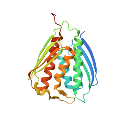 | |
Entity Groups | |||||
| Sequence Clusters | 30% Identity50% Identity70% Identity90% Identity95% Identity100% Identity | ||||
Sequence AnnotationsExpand | |||||
Reference Sequence | |||||
| Ligands 2 Unique | |||||
|---|---|---|---|---|---|
| ID | Chains | Name / Formula / InChI Key | 2D Diagram | 3D Interactions | |
| IYP Download:Ideal Coordinates CCD File | B [auth A] | (2R,3S)-2,3-dihydroxy-3-(1H-imidazol-5-yl)propyl dihydrogen phosphate C6 H11 N2 O6 P HFYBTHCYPKEDQQ-RITPCOANSA-N |  | ||
| MN Download:Ideal Coordinates CCD File | C [auth A], D [auth A], E [auth A] | MANGANESE (II) ION Mn WAEMQWOKJMHJLA-UHFFFAOYSA-N |  | ||
| Length ( Å ) | Angle ( ˚ ) |
|---|---|
| a = 112.514 | α = 90 |
| b = 112.514 | β = 90 |
| c = 112.514 | γ = 90 |
| Software Name | Purpose |
|---|---|
| MxCuBE | data collection |
| PHASER | phasing |
| REFMAC | refinement |
| HKL-2000 | data reduction |
| SCALEPACK | data scaling |


