4LN3 | pdb_00004ln3

The crystal structure of hemagglutinin from a H7N9 influenza virus (A/Shanghai/1/2013)


Experimental Data Snapshot

  • Method: X-RAY DIFFRACTION
  • Resolution: 2.65 Å
  • R-Value Free: 
    0.244 (Depositor), 0.244 (DCC) 
  • R-Value Work: 
    0.211 (Depositor), 0.213 (DCC) 
  • R-Value Observed: 
    0.212 (Depositor) 

wwPDB Validation 3D Report Full Report

Validation slider image for 4LN3

Ligand Structure Quality Assessment 


This is version 2.1 of the entry. See complete history

Literature

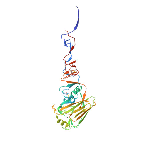
Structural Analysis of the Hemagglutinin from the Recent 2013 H7N9 Influenza Virus.

Yang, H.Carney, P.J.Chang, J.C.Villanueva, J.M.Stevens, J.

(2013) J Virol 87: 12433-12446

  • DOI: https://doi.org/10.1128/JVI.01854-13
  • Primary Citation Related Structures: 
    4LN3, 4LN4, 4LN6, 4LN8

  • PubMed Abstract: 

    In March 2013, the Chinese Center for Disease Control and Prevention reported human infections with an H7N9 influenza virus, and by 20 July 2013, the numbers of laboratory-confirmed cases had climbed to 134, including 43 fatalities and 127 hospitalizations. The newly emerging H7N9 viruses constitute an obvious public health concern because of the apparent severity of this outbreak. Here we focus on the hemagglutinins (HAs) of these viruses and assess their receptor binding phenotype in relation to previous HAs studied. Glycan microarray and kinetic analyses of recombinant A(H7N9) HAs were performed to compare the receptor binding profile of wild-type receptor binding site variants at position 217, a residue analogous to one of two positions known to switch avian to human receptor preference in H2N2 and H3N2 viruses. Two recombinant A(H7N9) HAs were structurally characterized, and a mutational study of the receptor binding site was performed to analyze important residues that can affect receptor preference and affinity. Results highlight a weak human receptor preference of the H7N9 HAs, suggesting that these viruses require further adaptation in order to adapt fully to humans.


  • Organizational Affiliation
    • Influenza Division, National Center for Immunization and Respiratory Diseases, Centers for Disease Control and Prevention, Atlanta, Georgia, USA.

Macromolecule Content 

  • Total Structure Weight: 341.7 kDa 
  • Atom Count: 23,076 
  • Modeled Residue Count: 2,889 
  • Deposited Residue Count: 3,036 
  • Unique protein chains: 2

Macromolecules

Find similar proteins by:|  3D Structure
Entity ID: 1
MoleculeChains  Sequence LengthOrganismDetailsImage
Hemagglutinin
A, C, E, G, I
A, C, E, G, I, K
325Influenza A virus (A/Shanghai/02/2013(H7N9))Mutation(s): 0 
UniProt
Find proteins for R4NN21 (Influenza A virus)
Explore R4NN21 
Go to UniProtKB:  R4NN21
Entity Groups
Sequence Clusters30% Identity50% Identity70% Identity90% Identity95% Identity100% Identity
UniProt GroupR4NN21
Glycosylation
Glycosylation Sites: 2
Sequence Annotations
Expand
Reference Sequence
Find similar proteins by:|  3D Structure
Entity ID: 2
MoleculeChains  Sequence LengthOrganismDetailsImage
Hemagglutinin
B, D, F, H, J
B, D, F, H, J, L
181Influenza A virus (A/Shanghai/02/2013(H7N9))Mutation(s): 0 
UniProt
Find proteins for R4NN21 (Influenza A virus)
Explore R4NN21 
Go to UniProtKB:  R4NN21
Entity Groups
Sequence Clusters30% Identity50% Identity70% Identity90% Identity95% Identity100% Identity
UniProt GroupR4NN21
Glycosylation
Glycosylation Sites: 1
Sequence Annotations
Expand
Reference Sequence

Oligosaccharides

Help  
Entity ID: 3
MoleculeChains Length2D Diagram GlycosylationD Interactions
beta-D-mannopyranose-(1-4)-2-acetamido-2-deoxy-beta-D-glucopyranose-(1-4)-2-acetamido-2-deoxy-beta-D-glucopyranose
M, N, O
3N-Glycosylation
Glycosylation Resources
GlyTouCan: G15407YE
GlyCosmos: G15407YE
GlyGen: G15407YE

Small Molecules

Ligands 1 Unique
IDChains Name / Formula / InChI Key2D Diagram3D Interactions
NAG

Query on NAG


Download:Ideal Coordinates CCD File
P [auth A]
Q [auth B]
R [auth D]
S [auth E]
T [auth F]
P [auth A],
Q [auth B],
R [auth D],
S [auth E],
T [auth F],
U [auth G],
V [auth H],
W [auth I],
X [auth J],
Y [auth K],
Z [auth L]
2-acetamido-2-deoxy-beta-D-glucopyranose
C8 H15 N O6
OVRNDRQMDRJTHS-FMDGEEDCSA-N

Experimental Data & Validation

Experimental Data

  • Method: X-RAY DIFFRACTION
  • Resolution: 2.65 Å
  • R-Value Free:  0.244 (Depositor), 0.244 (DCC) 
  • R-Value Work:  0.211 (Depositor), 0.213 (DCC) 
  • R-Value Observed: 0.212 (Depositor) 
Space Group: P 21 21 21
Unit Cell:
Length ( Å )Angle ( ˚ )
a = 154.297α = 90
b = 154.289β = 90
c = 154.933γ = 90
Software Package:
Software NamePurpose
HKL-2000data collection
PHASERphasing
REFMACrefinement
HKL-2000data reduction
HKL-2000data scaling

Structure Validation

View Full Validation Report


Ligand Structure Quality Assessment 


Entry History 

Deposition Data

Revision History  (Full details and data files)

  • Version 1.0: 2013-10-02
    Type: Initial release
  • Version 1.1: 2013-10-30
    Changes: Database references
  • Version 2.0: 2020-07-29
    Type: Remediation
    Reason: Carbohydrate remediation
    Changes: Advisory, Atomic model, Data collection, Derived calculations, Structure summary
  • Version 2.1: 2024-10-30
    Changes: Data collection, Database references, Refinement description, Structure summary