4LHD | pdb_00004lhd

Crystal structure of Synechocystis sp. PCC 6803 glycine decarboxylase (P-protein), holo form with pyridoxal-5'-phosphate and glycine, closed flexible loop


Experimental Data Snapshot

  • Method: X-RAY DIFFRACTION
  • Resolution: 1.80 Å
  • R-Value Free: 
    0.191 (Depositor), 0.189 (DCC) 
  • R-Value Work: 
    0.163 (Depositor), 0.161 (DCC) 
  • R-Value Observed: 
    0.164 (Depositor) 

Starting Model: experimental
View more details

wwPDB Validation 3D Report Full Report

Validation slider image for 4LHD

This is version 1.4 of the entry. See complete history

Literature

Structure of the Homodimeric Glycine Decarboxylase P-protein from Synechocystis sp. PCC 6803 Suggests a Mechanism for Redox Regulation.

Hasse, D.Andersson, E.Carlsson, G.Masloboy, A.Hagemann, M.Bauwe, H.Andersson, I.

(2013) J Biological Chem 288: 35333-35345

  • DOI: https://doi.org/10.1074/jbc.M113.509976
  • Primary Citation Related Structures: 
    4LGL, 4LHC, 4LHD

  • PubMed Abstract: 

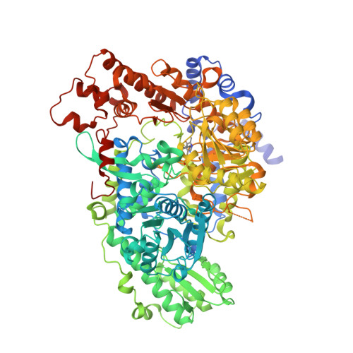
    Glycine decarboxylase, or P-protein, is a pyridoxal 5'-phosphate (PLP)-dependent enzyme in one-carbon metabolism of all organisms, in the glycine and serine catabolism of vertebrates, and in the photorespiratory pathway of oxygenic phototrophs. P-protein from the cyanobacterium Synechocystis sp. PCC 6803 is an α2 homodimer with high homology to eukaryotic P-proteins. The crystal structure of the apoenzyme shows the C terminus locked in a closed conformation by a disulfide bond between Cys(972) in the C terminus and Cys(353) located in the active site. The presence of the disulfide bridge isolates the active site from solvent and hinders the binding of PLP and glycine in the active site. Variants produced by substitution of Cys(972) and Cys(353) by Ser using site-directed mutagenesis have distinctly lower specific activities, supporting the crucial role of these highly conserved redox-sensitive amino acid residues for P-protein activity. Reduction of the 353-972 disulfide releases the C terminus and allows access to the active site. PLP and the substrate glycine bind in the active site of this reduced enzyme and appear to cause further conformational changes involving a flexible surface loop. The observation of the disulfide bond that acts to stabilize the closed form suggests a molecular mechanism for the redox-dependent activation of glycine decarboxylase observed earlier.


  • Organizational Affiliation
    • From the Department of Cell and Molecular Biology, Uppsala University, S-751 24 Uppsala, Sweden.

Macromolecule Content 

  • Total Structure Weight: 217.18 kDa 
  • Atom Count: 16,216 
  • Modeled Residue Count: 1,892 
  • Deposited Residue Count: 1,966 
  • Unique protein chains: 1

Macromolecules

Find similar proteins by:|  3D Structure
Entity ID: 1
MoleculeChains  Sequence LengthOrganismDetailsImage
Glycine dehydrogenase [decarboxylating]
A, B
983Synechocystis sp. PCC 6803 substr. KazusaMutation(s): 0 
Gene Names: gcvPslr0293
EC: 1.4.4.2
UniProt
Find proteins for P74416 (Synechocystis sp. (strain ATCC 27184 / PCC 6803 / Kazusa))
Explore P74416 
Go to UniProtKB:  P74416
Entity Groups
Sequence Clusters30% Identity50% Identity70% Identity90% Identity95% Identity100% Identity
UniProt GroupP74416
Sequence Annotations
Expand
Reference Sequence

Small Molecules

Ligands 4 Unique
IDChains Name / Formula / InChI Key2D Diagram3D Interactions
PO4

Query on PO4


Download:Ideal Coordinates CCD File
J [auth A],
K [auth A],
W [auth B],
X [auth B]
PHOSPHATE ION
O4 P
NBIIXXVUZAFLBC-UHFFFAOYSA-K
GLY

Query on GLY


Download:Ideal Coordinates CCD File
C [auth A]
D [auth A]
E [auth A]
F [auth A]
T [auth B]
C [auth A],
D [auth A],
E [auth A],
F [auth A],
T [auth B],
U [auth B]
GLYCINE
C2 H5 N O2
DHMQDGOQFOQNFH-UHFFFAOYSA-N
EDO

Query on EDO


Download:Ideal Coordinates CCD File
AA [auth B]
BA [auth B]
CA [auth B]
L [auth A]
M [auth A]
AA [auth B],
BA [auth B],
CA [auth B],
L [auth A],
M [auth A],
N [auth A],
O [auth A],
P [auth A],
Q [auth A],
R [auth A],
S [auth A],
Y [auth B],
Z [auth B]
1,2-ETHANEDIOL
C2 H6 O2
LYCAIKOWRPUZTN-UHFFFAOYSA-N
BCT

Query on BCT


Download:Ideal Coordinates CCD File
G [auth A],
H [auth A],
I [auth A],
V [auth B]
BICARBONATE ION
C H O3
BVKZGUZCCUSVTD-UHFFFAOYSA-M
Modified Residues  1 Unique
IDChains TypeFormula2D DiagramParent
LLP
Query on LLP
A, B
L-PEPTIDE LINKINGC14 H22 N3 O7 PLYS

Experimental Data & Validation

Experimental Data

  • Method: X-RAY DIFFRACTION
  • Resolution: 1.80 Å
  • R-Value Free:  0.191 (Depositor), 0.189 (DCC) 
  • R-Value Work:  0.163 (Depositor), 0.161 (DCC) 
  • R-Value Observed: 0.164 (Depositor) 
Space Group: P 3 2 1
Unit Cell:
Length ( Å )Angle ( ˚ )
a = 159.992α = 90
b = 159.992β = 90
c = 159.68γ = 120
Software Package:
Software NamePurpose
ADSCdata collection
PHASERphasing
PHENIXrefinement
DENZOdata reduction
SCALEPACKdata scaling

Structure Validation

View Full Validation Report


Entry History 

Deposition Data

Revision History  (Full details and data files)

  • Version 1.0: 2013-10-16
    Type: Initial release
  • Version 1.1: 2013-10-30
    Changes: Database references
  • Version 1.2: 2013-12-25
    Changes: Database references
  • Version 1.3: 2023-09-20
    Changes: Data collection, Database references, Derived calculations, Refinement description
  • Version 1.4: 2023-12-06
    Changes: Data collection