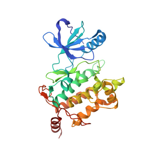
4L53

Crystal Structure of (1R,4R)-4-{4-[7-amino-2-(1,2,3-benzothiadiazol-7-yl)-3-chlorofuro[2,3-c]pyridin-4-yl]-1H-pyrazol-1-yl}cyclohexan-1-ol bound to TAK1-TAB1


Experimental Data Snapshot

  • Method: X-RAY DIFFRACTION
  • Resolution: 2.55 Å
  • R-Value Free: 0.238 
  • R-Value Work: 0.209 
  • R-Value Observed: 0.211 

wwPDB Validation   3D Report Full Report


Ligand Structure Quality Assessment 


This is version 1.5 of the entry. See complete history


Literature

Discovery of 7-aminofuro[2,3-c]pyridine inhibitors of TAK1: Optimization of kinase selectivity and pharmacokinetics.

Hornberger, K.R.Chen, X.Crew, A.P.Kleinberg, A.Ma, L.Mulvihill, M.J.Wang, J.Wilde, V.L.Albertella, M.Bittner, M.Cooke, A.Kadhim, S.Kahler, J.Maresca, P.May, E.Meyn, P.Romashko, D.Tokar, B.Turton, R.

(2013) Bioorg Med Chem Lett 23: 4511-4516

  • DOI: https://doi.org/10.1016/j.bmcl.2013.06.054
  • Primary Citation of Related Structures:  
    4L53

  • PubMed Abstract: 

    The kinase selectivity and pharmacokinetic optimization of a series of 7-aminofuro[2,3-c]pyridine inhibitors of TAK1 is described. The intersection of insights from molecular modeling, computational prediction of metabolic sites, and in vitro metabolite identification studies resulted in a simple and unique solution to both of these problems. These efforts culminated in the discovery of compound 13a, a potent, relatively selective inhibitor of TAK1 with good pharmacokinetic properties in mice, which was active in an in vivo model of ovarian cancer.


  • Organizational Affiliation

    OSI Pharmaceuticals LLC, 1 Bioscience Park Drive, Farmingdale, NY 11735, USA. keith.hornberger@boehringer-ingelheim.com


Macromolecules
Find similar proteins by:  (by identity cutoff)  |  3D Structure
Entity ID: 1
MoleculeChains Sequence LengthOrganismDetailsImage
Mitogen-activated protein kinase kinase kinase 7, TGF-beta-activated kinase 1 and MAP3K7-binding protein 1 chimera307Homo sapiensMutation(s): 0 
Gene Names: MAP3K7TAK1MAP3K7IP1TAB1
EC: 2.7.11.25
UniProt & NIH Common Fund Data Resources
Find proteins for Q15750 (Homo sapiens)
Explore Q15750 
Go to UniProtKB:  Q15750
PHAROS:  Q15750
GTEx:  ENSG00000100324 
Find proteins for O43318 (Homo sapiens)
Explore O43318 
Go to UniProtKB:  O43318
PHAROS:  O43318
GTEx:  ENSG00000135341 
Entity Groups  
Sequence Clusters30% Identity50% Identity70% Identity90% Identity95% Identity100% Identity
UniProt GroupsO43318Q15750
Sequence Annotations
Expand
  • Reference Sequence
Small Molecules
Binding Affinity Annotations 
IDSourceBinding Affinity
1UO PDBBind:  4L53 IC50: 28 (nM) from 1 assay(s)
Experimental Data & Validation

Experimental Data

  • Method: X-RAY DIFFRACTION
  • Resolution: 2.55 Å
  • R-Value Free: 0.238 
  • R-Value Work: 0.209 
  • R-Value Observed: 0.211 
  • Space Group: I 2 2 2
Unit Cell:
Length ( Å )Angle ( ˚ )
a = 58.568α = 90
b = 134.53β = 90
c = 143.689γ = 90
Software Package:
Software NamePurpose
MOLREPphasing
REFMACrefinement
XDSdata reduction
XSCALEdata scaling

Structure Validation

View Full Validation Report


Ligand Structure Quality Assessment 


Entry History 

Deposition Data

Revision History  (Full details and data files)

  • Version 1.0: 2013-07-03
    Type: Initial release
  • Version 1.1: 2013-07-31
    Changes: Database references
  • Version 1.2: 2013-08-14
    Changes: Database references
  • Version 1.3: 2017-07-26
    Changes: Advisory, Refinement description, Source and taxonomy
  • Version 1.4: 2017-11-15
    Changes: Refinement description
  • Version 1.5: 2024-02-28
    Changes: Advisory, Data collection, Database references, Derived calculations