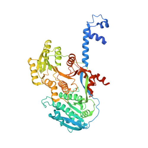
4L4X

An A2-type ketoreductase from a modular polyketide synthase


Experimental Data Snapshot

  • Method: X-RAY DIFFRACTION
  • Resolution: 2.55 Å
  • R-Value Free: 0.280 
  • R-Value Work: 0.227 
  • R-Value Observed: 0.230 

Starting Model: experimental
View more details

wwPDB Validation   3D Report Full Report


Ligand Structure Quality Assessment 


This is version 1.2 of the entry. See complete history


Literature

Structural Studies of an A2-Type Modular Polyketide Synthase Ketoreductase Reveal Features Controlling alpha-Substituent Stereochemistry.

Zheng, J.Piasecki, S.K.Keatinge-Clay, A.T.

(2013) ACS Chem Biol 8: 1964-1971

  • DOI: https://doi.org/10.1021/cb400161g
  • Primary Citation of Related Structures:  
    4L4X

  • PubMed Abstract: 

    Modular polyketide synthase ketoreductases often set two stereocenters when reducing intermediates in the biosynthesis of a complex polyketide. Here we report the 2.55-Å resolution structure of an A2-type ketoreductase from the 11th module of the amphotericin polyketide synthase that sets a combination of l-α-methyl and l-β-hydroxyl stereochemistries and represents the final catalytically competent ketoreductase type to be structurally elucidated. Through structure-guided mutagenesis a double mutant of an A1-type ketoreductase was generated that functions as an A2-type ketoreductase on a diketide substrate analogue, setting an α-alkyl substituent in an l-orientation rather than in the d-orientation set by the unmutated ketoreductase. When the activity of the double mutant was examined in the context of an engineered triketide lactone synthase, the anticipated triketide lactone was not produced even though the ketoreductase-containing module still reduced the diketide substrate analogue as expected. These findings suggest that re-engineered ketoreductases may be catalytically outcompeted within engineered polyketide synthase assembly lines.


  • Organizational Affiliation

    Department of Chemistry and Biochemistry and ‡Institute for Cellular and Molecular Biology, The University of Texas at Austin , Austin, Texas 78712, United States.


Macromolecules
Find similar proteins by:  (by identity cutoff)  |  3D Structure
Entity ID: 1
MoleculeChains Sequence LengthOrganismDetailsImage
AmphI580Streptomyces nodosusMutation(s): 0 
Gene Names: amphI
EC: 1.1.1.100
UniProt
Find proteins for Q93NX9 (Streptomyces nodosus)
Explore Q93NX9 
Go to UniProtKB:  Q93NX9
Entity Groups  
Sequence Clusters30% Identity50% Identity70% Identity90% Identity95% Identity100% Identity
UniProt GroupQ93NX9
Sequence Annotations
Expand
  • Reference Sequence
Small Molecules
Ligands 1 Unique
IDChains Name / Formula / InChI Key2D Diagram3D Interactions
NDP
Query on NDP

Download Ideal Coordinates CCD File 
B [auth A]NADPH DIHYDRO-NICOTINAMIDE-ADENINE-DINUCLEOTIDE PHOSPHATE
C21 H30 N7 O17 P3
ACFIXJIJDZMPPO-NNYOXOHSSA-N
Experimental Data & Validation

Experimental Data

  • Method: X-RAY DIFFRACTION
  • Resolution: 2.55 Å
  • R-Value Free: 0.280 
  • R-Value Work: 0.227 
  • R-Value Observed: 0.230 
  • Space Group: C 2 2 21
Unit Cell:
Length ( Å )Angle ( ˚ )
a = 45.139α = 90
b = 133.513β = 90
c = 185.137γ = 90
Software Package:
Software NamePurpose
PHASESphasing
REFMACrefinement
HKL-2000data reduction
HKL-2000data scaling

Structure Validation

View Full Validation Report


Ligand Structure Quality Assessment 


Entry History 

Deposition Data

Revision History  (Full details and data files)

  • Version 1.0: 2013-06-19
    Type: Initial release
  • Version 1.1: 2013-10-09
    Changes: Database references
  • Version 1.2: 2023-09-20
    Changes: Data collection, Database references, Derived calculations, Refinement description