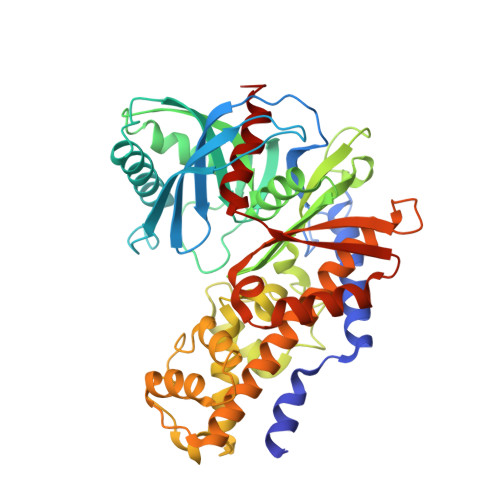
4L3Q | pdb_00004l3q

Crystal structure of glucokinase-activator complex


Experimental Data Snapshot

  • Method: X-RAY DIFFRACTION
  • Resolution: 2.70 Å
  • R-Value Free: 
    0.304 (Depositor), 0.290 (DCC) 
  • R-Value Work: 
    0.254 (Depositor), 0.240 (DCC) 
  • R-Value Observed: 
    0.259 (Depositor) 

Starting Model: experimental
View more details

wwPDB Validation   3D Report Full Report


Ligand Structure Quality Assessment 

Created with Raphaël 2.3.0Worse 01 BetterLigand structure goodness of fit to experimental dataBest fitted 926Click on this verticalbar to view detailsBest fitted GLCClick on this verticalbar to view details

This is version 1.3 of the entry. See complete history


Literature

Pyrimidone-based series of glucokinase activators with alternative donor-acceptor motif.

Filipski, K.J.Guzman-Perez, A.Bian, J.Perreault, C.Aspnes, G.E.Didiuk, M.T.Dow, R.L.Hank, R.F.Jones, C.S.Maguire, R.J.Tu, M.Zeng, D.Liu, S.Knafels, J.D.Litchfield, J.Atkinson, K.Derksen, D.R.Bourbonais, F.Gajiwala, K.S.Hickey, M.Johnson, T.O.Humphries, P.S.Pfefferkorn, J.A.

(2013) Bioorg Med Chem Lett 23: 4571-4578

  • DOI: https://doi.org/10.1016/j.bmcl.2013.06.036
  • Primary Citation of Related Structures:  
    4L3Q

  • PubMed Abstract: 

    Glucokinase activators are a class of experimental agents under investigation as a therapy for Type 2 diabetes mellitus. An X-ray crystal structure of a modestly potent agent revealed the potential to substitute the common heterocyclic amide donor-acceptor motif for a pyridone moiety. We have successfully demonstrated that both pyridone and pyrimidone heterocycles can be used as a potent donor-acceptor substituent. Several sub-micromolar analogs that possess the desired partial activator profile were synthesized and characterized. Unfortunately, the most potent activators suffered from sub-optimal pharmacokinetic properties. Nonetheless, these donor-acceptor motifs may find utility in other glucokinase activator series or beyond.


  • Organizational Affiliation

    Cardiovascular, Metabolic, and Endocrine Diseases Chemistry, Pfizer Worldwide Research and Development, 620 Memorial Dr, Cambridge, MA 02139, USA. kevin.filipski@pfizer.com


Macromolecules
Find similar proteins by:  (by identity cutoff)  |  3D Structure
Entity ID: 1
MoleculeChains Sequence LengthOrganismDetailsImage
Glucokinase457Homo sapiensMutation(s): 0 
Gene Names: GCK
EC: 2.7.1.2 (PDB Primary Data), 2.7.1.1 (UniProt)
UniProt & NIH Common Fund Data Resources
Find proteins for P35557 (Homo sapiens)
Explore P35557 
Go to UniProtKB:  P35557
PHAROS:  P35557
GTEx:  ENSG00000106633 
Entity Groups  
Sequence Clusters30% Identity50% Identity70% Identity90% Identity95% Identity100% Identity
UniProt GroupP35557
Sequence Annotations
Expand
  • Reference Sequence
Small Molecules
Ligands 2 Unique
IDChains Name / Formula / InChI Key2D Diagram3D Interactions
926
Query on 926

Download Ideal Coordinates CCD File 
C [auth A]6-{3-[(1-methyl-1H-imidazol-2-yl)sulfanyl]phenyl}pyridin-2(1H)-one
C15 H13 N3 O S
YYWBGPHQPMDZEM-UHFFFAOYSA-N
GLC
Query on GLC

Download Ideal Coordinates CCD File 
B [auth A]alpha-D-glucopyranose
C6 H12 O6
WQZGKKKJIJFFOK-DVKNGEFBSA-N
Binding Affinity Annotations 
IDSourceBinding Affinity
926 BindingDB:  4L3Q EC50: 2000 (nM) from 1 assay(s)
Experimental Data & Validation

Experimental Data

  • Method: X-RAY DIFFRACTION
  • Resolution: 2.70 Å
  • R-Value Free:  0.304 (Depositor), 0.290 (DCC) 
  • R-Value Work:  0.254 (Depositor), 0.240 (DCC) 
  • R-Value Observed: 0.259 (Depositor) 
Space Group: P 65 2 2
Unit Cell:
Length ( Å )Angle ( ˚ )
a = 79.512α = 90
b = 79.512β = 90
c = 321.131γ = 120
Software Package:
Software NamePurpose
MAR345dtbdata collection
CNXrefinement
HKL-2000data reduction
SCALEPACKdata scaling
CNXphasing

Structure Validation

View Full Validation Report


Ligand Structure Quality Assessment 

Created with Raphaël 2.3.0Worse 01 BetterLigand structure goodness of fit to experimental dataBest fitted 926Click on this verticalbar to view detailsBest fitted GLCClick on this verticalbar to view details

Entry History 

Deposition Data

Revision History  (Full details and data files)

  • Version 1.0: 2013-07-24
    Type: Initial release
  • Version 1.1: 2013-08-07
    Changes: Database references
  • Version 1.2: 2020-07-29
    Type: Remediation
    Reason: Carbohydrate remediation
    Changes: Data collection, Database references, Derived calculations, Structure summary
  • Version 1.3: 2023-09-20
    Changes: Data collection, Database references, Refinement description, Structure summary