4KWF | pdb_00004kwf

Crystal Structure Analysis of ALDH2+ALDiB33


Experimental Data Snapshot

  • Method: X-RAY DIFFRACTION
  • Resolution: 2.31 Å
  • R-Value Free: 
    0.296 (Depositor), 0.293 (DCC) 
  • R-Value Work: 
    0.239 (Depositor), 0.238 (DCC) 
  • R-Value Observed: 
    0.242 (Depositor) 

wwPDB Validation 3D Report Full Report

Validation slider image for 4KWF

Ligand Structure Quality Assessment 


This is version 1.2 of the entry. See complete history

Literature

Development of selective inhibitors for aldehyde dehydrogenases based on substituted indole-2,3-diones.

Kimble-Hill, A.C.Parajuli, B.Chen, C.H.Mochly-Rosen, D.Hurley, T.D.

(2014) J Med Chem 57: 714-722

  • DOI: https://doi.org/10.1021/jm401377v
  • Primary Citation Related Structures: 
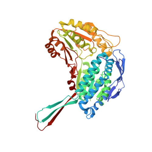
    4KWF, 4KWG, 4L1O

  • PubMed Abstract: 

    Aldehyde dehydrogenases (ALDH) participate in multiple metabolic pathways and have been indicated to play a role in several cancerous disease states. Our laboratory is interested in developing novel and selective ALDH inhibitors. We looked to further work recently published by developing a class of isoenzyme-selective inhibitors using similar indole-2,3-diones that exhibit differential inhibition of ALDH1A1, ALDH2, and ALDH3A1. Kinetic and X-ray crystallography data suggest that these inhibitors are competitive against aldehyde binding, forming direct interactions with active-site cysteine residues. The selectivity is precise in that these compounds appear to interact directly with the catalytic nucleophile, Cys243, in ALDH3A1 but not in ALDH2. In ALDH2, the 3-keto group is surrounded by the adjacent Cys301/303. Surprisingly, the orientation of the interaction changes depending on the nature of the substitutions on the basic indole ring structure and correlates well with the observed structure-activity relationships for each ALDH isoenzyme.


  • Organizational Affiliation
    • Department of Biochemistry & Molecular Biology, Indiana University School of Medicine , MS4053, 635 Barnhill Drive, Indianapolis, Indiana 46202, United States.

Macromolecule Content 

  • Total Structure Weight: 433.8 kDa 
  • Atom Count: 30,712 
  • Modeled Residue Count: 3,952 
  • Deposited Residue Count: 3,952 
  • Unique protein chains: 1

Macromolecules

Find similar proteins by:|  3D Structure
Entity ID: 1
MoleculeChains  Sequence LengthOrganismDetailsImage
Aldehyde dehydrogenase, mitochondrial
A, B, C, D, E
A, B, C, D, E, F, G, H
494Homo sapiensMutation(s): 0 
Gene Names: ALDH2ALDM
EC: 1.2.1.3
UniProt & NIH Common Fund Data Resources
Find proteins for P05091 (Homo sapiens)
Explore P05091 
Go to UniProtKB:  P05091
PHAROS:  P05091
GTEx:  ENSG00000111275 
Entity Groups
Sequence Clusters30% Identity50% Identity70% Identity90% Identity95% Identity100% Identity
UniProt GroupP05091
Sequence Annotations
Expand
Reference Sequence

Small Molecules

Ligands 4 Unique
IDChains Name / Formula / InChI Key2D Diagram3D Interactions
3AK

Query on 3AK


Download:Ideal Coordinates CCD File
FA [auth H],
I [auth A],
N [auth B],
W [auth E]
1-benzyl-1H-indole-2,3-dione
C15 H11 N O2
SIISFRLGYDVIRG-UHFFFAOYSA-N
EDO

Query on EDO


Download:Ideal Coordinates CCD File
BA [auth F]
HA [auth H]
K [auth A]
L [auth A]
R [auth C]
BA [auth F],
HA [auth H],
K [auth A],
L [auth A],
R [auth C],
S [auth C],
Y [auth E],
Z [auth E]
1,2-ETHANEDIOL
C2 H6 O2
LYCAIKOWRPUZTN-UHFFFAOYSA-N
GAI

Query on GAI


Download:Ideal Coordinates CCD File
CA [auth F]
EA [auth G]
IA [auth H]
M [auth A]
P [auth B]
CA [auth F],
EA [auth G],
IA [auth H],
M [auth A],
P [auth B],
T [auth C],
V [auth D]
GUANIDINE
C H5 N3
ZRALSGWEFCBTJO-UHFFFAOYSA-N
NA

Query on NA


Download:Ideal Coordinates CCD File
AA [auth F]
DA [auth G]
GA [auth H]
J [auth A]
O [auth B]
AA [auth F],
DA [auth G],
GA [auth H],
J [auth A],
O [auth B],
Q [auth C],
U [auth D],
X [auth E]
SODIUM ION
Na
FKNQFGJONOIPTF-UHFFFAOYSA-N
Binding Affinity Annotations 
IDSourceBinding Affinity
3AK BindingDB:  4KWF Ki: min: 1.50e+4, max: 3.40e+4 (nM) from 2 assay(s)
IC50: 8.20e+4 (nM) from 1 assay(s)

Experimental Data & Validation

Experimental Data

  • Method: X-RAY DIFFRACTION
  • Resolution: 2.31 Å
  • R-Value Free:  0.296 (Depositor), 0.293 (DCC) 
  • R-Value Work:  0.239 (Depositor), 0.238 (DCC) 
  • R-Value Observed: 0.242 (Depositor) 
Space Group: P 1 21 1
Unit Cell:
Length ( Å )Angle ( ˚ )
a = 102.293α = 90
b = 177.092β = 94.39
c = 102.551γ = 90
Software Package:
Software NamePurpose
DENZOdata reduction
SCALEPACKdata scaling
REFMACrefinement
PDB_EXTRACTdata extraction
HKL-3000data collection
HKL-3000data reduction
HKL-3000data scaling
REFMACphasing

Structure Validation

View Full Validation Report


Ligand Structure Quality Assessment 


Entry History 

Deposition Data

Revision History  (Full details and data files)

  • Version 1.0: 2014-04-09
    Type: Initial release
  • Version 1.1: 2017-11-15
    Changes: Refinement description
  • Version 1.2: 2024-11-06
    Changes: Data collection, Database references, Derived calculations, Structure summary