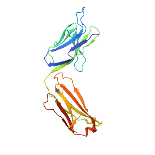
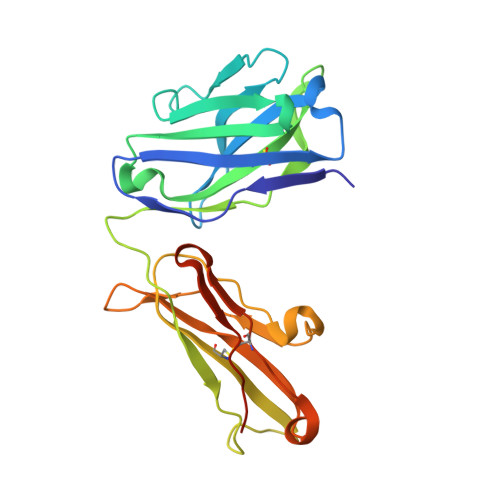
4KUZ

Crystal structure of anti-emmprin antibody 4A5 Fab in trigonal form


Experimental Data Snapshot

  • Method: X-RAY DIFFRACTION
  • Resolution: 2.70 Å
  • R-Value Free: 0.268 
  • R-Value Work: 0.230 
  • R-Value Observed: 0.233 

Starting Models: experimental
View more details

wwPDB Validation   3D Report Full Report


This is version 1.3 of the entry. See complete history


Literature

Structural evidence for a constrained conformation of short CDR-L3 in antibodies.

Teplyakov, A.Obmolova, G.Malia, T.J.Luo, J.Gilliland, G.L.

(2014) Proteins 82: 1679-1683

  • DOI: https://doi.org/10.1002/prot.24522
  • Primary Citation of Related Structures:  
    4KQ3, 4KUZ

  • PubMed Abstract: 

    Three Fab structures used as targets in the Antibody Modeling Assessment presented a challenge for modeling CDR-L3 due to deviations from the most typical canonical structure. In all three antibodies CDR-L3 has eight residues, which is one residue shorter than usual, and has a conformation that is rarely observed in crystal structures. We analyzed the sequence and structural determinants of this conformation and found that the "short" CDR-L3 is remarkably rigid and retains the conformation in the interactions with antigens and neighboring CDRs.


  • Organizational Affiliation

    Janssen Research & Development, LLC, Spring House, Pennsylvania, 19477.


Macromolecules
Find similar proteins by:  (by identity cutoff)  |  3D Structure
Entity ID: 1
MoleculeChains Sequence LengthOrganismDetailsImage
antibody 4A5 light chainA [auth L]219Mus musculusHomo sapiens
This entity is chimeric
Mutation(s): 0 
Entity Groups  
Sequence Clusters30% Identity50% Identity70% Identity90% Identity95% Identity100% Identity
Sequence Annotations
Expand
  • Reference Sequence
Find similar proteins by:  (by identity cutoff)  |  3D Structure
Entity ID: 2
MoleculeChains Sequence LengthOrganismDetailsImage
antibody 4A5 heavy chainB [auth H]229Mus musculusHomo sapiens
This entity is chimeric
Mutation(s): 0 
Entity Groups  
Sequence Clusters30% Identity50% Identity70% Identity90% Identity95% Identity100% Identity
Sequence Annotations
Expand
  • Reference Sequence
Experimental Data & Validation

Experimental Data

  • Method: X-RAY DIFFRACTION
  • Resolution: 2.70 Å
  • R-Value Free: 0.268 
  • R-Value Work: 0.230 
  • R-Value Observed: 0.233 
  • Space Group: P 31 2 1
Unit Cell:
Length ( Å )Angle ( ˚ )
a = 126.43α = 90
b = 126.43β = 90
c = 97.15γ = 120
Software Package:
Software NamePurpose
PHENIXrefinement
AMoREphasing
REFMACrefinement
XDSdata reduction
XDSdata scaling
CrystalCleardata collection

Structure Validation

View Full Validation Report


Entry History 

Deposition Data

Revision History  (Full details and data files)

  • Version 1.0: 2014-03-26
    Type: Initial release
  • Version 1.1: 2014-08-06
    Changes: Database references
  • Version 1.2: 2019-07-17
    Changes: Data collection, Refinement description
  • Version 1.3: 2023-09-20
    Changes: Data collection, Database references, Derived calculations, Refinement description