


Crystal structure of a TrmH family tRNA methyltransferase bound to S-adenosyl homocysteine
Edwards, T.E., Fairman, J.W., Seattle Structural Genomics Center for Infectious Disease (SSGCID)To be published.
Experimental Data Snapshot
Entity ID: 1 | |||||
|---|---|---|---|---|---|
| Molecule | Chains | Sequence Length | Organism | Details | Image |
| tRNA (cytidine(34)-2'-O)-methyltransferase | 160 | Burkholderia pseudomallei 1710b | Mutation(s): 0 Gene Names: BURPS1710b_0668, trmL EC: 2.1.1.207 | 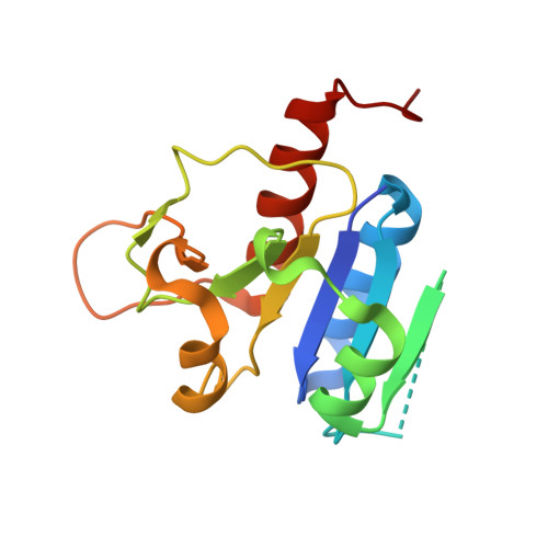 | |
UniProt | |||||
Entity Groups | |||||
| Sequence Clusters | 30% Identity50% Identity70% Identity90% Identity95% Identity100% Identity | ||||
| UniProt Group | Q3JWH1 | ||||
Sequence AnnotationsExpand | |||||
Reference Sequence | |||||
| Ligands 2 Unique | |||||
|---|---|---|---|---|---|
| ID | Chains | Name / Formula / InChI Key | 2D Diagram | 3D Interactions | |
| SAH Download:Ideal Coordinates CCD File | I [auth A], L [auth C], N [auth E], Q [auth G] | S-ADENOSYL-L-HOMOCYSTEINE C14 H20 N6 O5 S ZJUKTBDSGOFHSH-WFMPWKQPSA-N |  | ||
| CL Download:Ideal Coordinates CCD File | J [auth A] K [auth B] M [auth C] O [auth E] P [auth F] | CHLORIDE ION Cl VEXZGXHMUGYJMC-UHFFFAOYSA-M |  | ||
| Length ( Å ) | Angle ( ˚ ) |
|---|---|
| a = 52.6 | α = 90 |
| b = 149.71 | β = 90.49 |
| c = 80.3 | γ = 90 |
| Software Name | Purpose |
|---|---|
| XSCALE | data scaling |
| PHASER | phasing |
| REFMAC | refinement |
| PDB_EXTRACT | data extraction |
| XDS | data reduction |


