The Structure of the Neisserial Lipooligosaccharide Phosphoethanolamine Transferase A (LptA) Required for Resistance to Polymyxin.
Wanty, C., Anandan, A., Piek, S., Walshe, J., Ganguly, J., Carlson, R.W., Stubbs, K.A., Kahler, C.M., Vrielink, A.(2013) J Mol Biology 425: 3389-3402
- PubMed: 23810904 Search on PubMed
- DOI: https://doi.org/10.1016/j.jmb.2013.06.029
- Primary Citation Related Structures:
4KAV, 4KAY - PubMed Abstract:
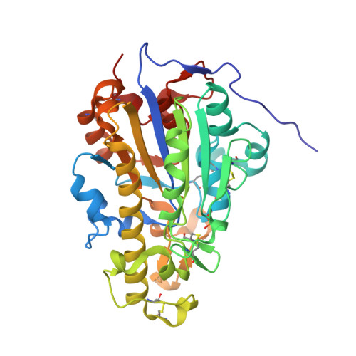
Gram-negative bacteria possess an outer membrane envelope consisting of an outer leaflet of lipopolysaccharides, also called endotoxins, which protect the pathogen from antimicrobial peptides and have multifaceted roles in virulence. Lipopolysaccharide consists of a glycan moiety attached to lipid A, embedded in the outer membrane. Modification of the lipid A headgroups by phosphoethanolamine (PEA) or 4-amino-arabinose residues increases resistance to the cationic cyclic polypeptide antibiotic, polymyxin. Lipid A PEA transferases are members of the YhjW/YjdB/YijP superfamily and usually consist of a transmembrane domain anchoring the enzyme to the periplasmic face of the cytoplasmic membrane attached to a soluble catalytic domain. The crystal structure of the soluble domain of the protein of the lipid A PEA transferase from Neisseria meningitidis has been determined crystallographically and refined to 1.4Å resolution. The structure reveals a core hydrolase fold similar to that of alkaline phosphatase. Loop regions in the structure differ, presumably to enable interaction with the membrane-localized substrates and to provide substrate specificity. A phosphorylated form of the putative nucleophile, Thr280, is observed. Metal ions present in the active site are coordinated to Thr280 and to residues conserved among the family of transferases. The structure reveals the protein components needed for the transferase chemistry; however, substrate-binding regions are not evident and are likely to reside in the transmembrane domain of the protein.
- School of Chemistry and Biochemistry, University of Western Australia, 35 Stirling Highway, Crawley, WA 6009, Australia.
Organizational Affiliation: