4K3I | pdb_00004k3i

Crystal Structure of the Quinol Form of Methylamine Dehydrogenase in Complex with the Diferrous Form of MauG, C2 Space Group


Experimental Data Snapshot

  • Method: X-RAY DIFFRACTION
  • Resolution: 2.00 Å
  • R-Value Free: 
    0.190 (Depositor), 0.198 (DCC) 
  • R-Value Work: 
    0.144 (Depositor), 0.155 (DCC) 
  • R-Value Observed: 
    0.146 (Depositor) 

Starting Model: experimental
View more details

wwPDB Validation 3D Report Full Report

Validation slider image for 4K3I

Ligand Structure Quality Assessment 


This is version 1.4 of the entry. See complete history

Literature

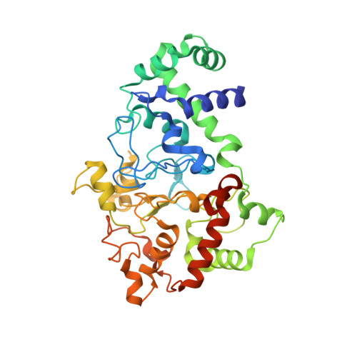
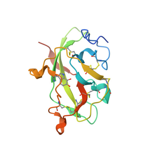
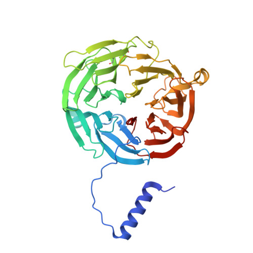
Structures of MauG in complex with quinol and quinone MADH.

Yukl, E.T.Jensen, L.M.Davidson, V.L.Wilmot, C.M.

(2013) Acta Crystallogr Sect F Struct Biol Cryst Commun 69: 738-743

  • DOI: https://doi.org/10.1107/S1744309113016539
  • Primary Citation Related Structures: 
    4K3I

  • PubMed Abstract: 

    MauG has been cocrystallized with methylamine dehydrogenase (MADH) with its TTQ cofactor in the o-quinol (TTQOQ) and quinone (TTQOX) forms and the structures of the resulting complexes have been solved. The TTQOQ structure crystallized in either space group P21 or C2, while the TTQOX structure crystallized in space group P1. The previously solved structure of MauG in complex with MADH bearing an incompletely formed TTQ cofactor (preMADH) also crystallized in space group P1, although with different unit-cell parameters. Despite the changes in crystal form, the structures are virtually identical, with only very minor changes at the protein-protein interface. The relevance of these structures with respect to the measured changes in affinity between MauG and various forms of MADH is discussed.


  • Organizational Affiliation
    • Department of Biochemistry, Molecular Biology and Biophysics, University of Minnesota, 321 Church Street SE, Minneapolis, MN 55455, USA.

Macromolecule Content 

  • Total Structure Weight: 199.97 kDa 
  • Atom Count: 15,344 
  • Modeled Residue Count: 1,713 
  • Deposited Residue Count: 1,790 
  • Unique protein chains: 3

Macromolecules

Find similar proteins by:|  3D Structure
Entity ID: 1
MoleculeChains  Sequence LengthOrganismDetailsImage
Methylamine utilization protein MauG
A, B
373Paracoccus denitrificans PD1222Mutation(s): 0 
Gene Names: mauGPden_4736
EC: 1
UniProt
Find proteins for Q51658 (Paracoccus denitrificans (strain Pd 1222))
Explore Q51658 
Go to UniProtKB:  Q51658
Entity Groups
Sequence Clusters30% Identity50% Identity70% Identity90% Identity95% Identity100% Identity
UniProt GroupQ51658
Sequence Annotations
Expand
Reference Sequence
Find similar proteins by:|  3D Structure
Entity ID: 2
MoleculeChains  Sequence LengthOrganismDetailsImage
Methylamine dehydrogenase light chain
C, E
137Paracoccus denitrificansMutation(s): 0 
Gene Names: mauA
EC: 1.4.9.1
UniProt
Find proteins for P22619 (Paracoccus denitrificans)
Explore P22619 
Go to UniProtKB:  P22619
Entity Groups
Sequence Clusters30% Identity50% Identity70% Identity90% Identity95% Identity100% Identity
UniProt GroupP22619
Sequence Annotations
Expand
Reference Sequence
Find similar proteins by:|  3D Structure
Entity ID: 3
MoleculeChains  Sequence LengthOrganismDetailsImage
Methylamine dehydrogenase heavy chain
D, F
385Paracoccus denitrificans PD1222Mutation(s): 0 
Gene Names: Pden_4730
EC: 1.4.99.3 (PDB Primary Data), 1.4.9.1 (UniProt)
UniProt
Find proteins for A1BB97 (Paracoccus denitrificans (strain Pd 1222))
Explore A1BB97 
Go to UniProtKB:  A1BB97
Entity Groups
Sequence Clusters30% Identity50% Identity70% Identity90% Identity95% Identity100% Identity
UniProt GroupA1BB97
Sequence Annotations
Expand
Reference Sequence

Small Molecules

Ligands 5 Unique
IDChains Name / Formula / InChI Key2D Diagram3D Interactions
HEC

Query on HEC


Download:Ideal Coordinates CCD File
H [auth A],
I [auth A],
P [auth B],
Q [auth B]
HEME C
C34 H34 Fe N4 O4
HXQIYSLZKNYNMH-LJNAALQVSA-N
EDO

Query on EDO


Download:Ideal Coordinates CCD File
K [auth A],
L [auth A],
N [auth A],
S [auth B]
1,2-ETHANEDIOL
C2 H6 O2
LYCAIKOWRPUZTN-UHFFFAOYSA-N
ACT

Query on ACT


Download:Ideal Coordinates CCD File
U [auth D]ACETATE ION
C2 H3 O2
QTBSBXVTEAMEQO-UHFFFAOYSA-M
CA

Query on CA


Download:Ideal Coordinates CCD File
G [auth A],
O [auth B]
CALCIUM ION
Ca
BHPQYMZQTOCNFJ-UHFFFAOYSA-N
NA

Query on NA


Download:Ideal Coordinates CCD File
J [auth A],
M [auth A],
R [auth B],
T [auth B]
SODIUM ION
Na
FKNQFGJONOIPTF-UHFFFAOYSA-N
Modified Residues  1 Unique
IDChains TypeFormula2D DiagramParent
TOQ
Query on TOQ
C, E
L-PEPTIDE LINKINGC11 H12 N2 O4TRP

Experimental Data & Validation

Experimental Data

  • Method: X-RAY DIFFRACTION
  • Resolution: 2.00 Å
  • R-Value Free:  0.190 (Depositor), 0.198 (DCC) 
  • R-Value Work:  0.144 (Depositor), 0.155 (DCC) 
  • R-Value Observed: 0.146 (Depositor) 
Space Group: C 1 2 1
Unit Cell:
Length ( Å )Angle ( ˚ )
a = 346.356α = 90
b = 55.558β = 106.55
c = 112.548γ = 90
Software Package:
Software NamePurpose
CBASSdata collection
PHASERphasing
REFMACrefinement
HKL-2000data reduction
HKL-2000data scaling

Structure Validation

View Full Validation Report


Ligand Structure Quality Assessment 


Entry History 

Deposition Data

Revision History  (Full details and data files)

  • Version 1.0: 2013-07-10
    Type: Initial release
  • Version 1.1: 2013-08-28
    Changes: Database references
  • Version 1.2: 2023-09-20
    Changes: Data collection, Database references, Derived calculations, Refinement description
  • Version 1.3: 2023-12-06
    Changes: Data collection, Derived calculations
  • Version 1.4: 2024-11-27
    Changes: Structure summary