4K3D | pdb_00004k3d

Crystal structure of bovine antibody BLV1H12 with ultralong CDR H3


Experimental Data Snapshot

  • Method: X-RAY DIFFRACTION
  • Resolution: 1.85 Å
  • R-Value Free: 
    0.210 (Depositor), 0.216 (DCC) 
  • R-Value Work: 
    0.182 (Depositor), 0.189 (DCC) 
  • R-Value Observed: 
    0.184 (Depositor) 

Starting Models: experimental
View more details

wwPDB Validation 3D Report Full Report

Validation slider image for 4K3D

This is version 2.1 of the entry. See complete history

Literature

Reshaping antibody diversity.

Wang, F.Ekiert, D.C.Ahmad, I.Yu, W.Zhang, Y.Bazirgan, O.Torkamani, A.Raudsepp, T.Mwangi, W.Criscitiello, M.F.Wilson, I.A.Schultz, P.G.Smider, V.V.

(2013) Cell 153: 1379-1393

  • DOI: https://doi.org/10.1016/j.cell.2013.04.049
  • Primary Citation Related Structures: 
    4K3D, 4K3E

  • PubMed Abstract: 

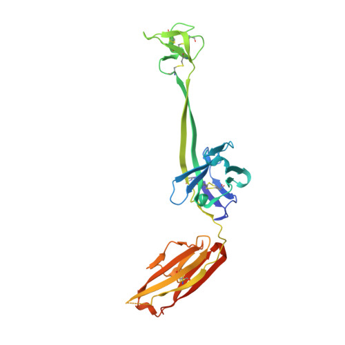
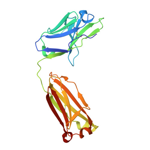
    Some species mount a robust antibody response despite having limited genome-encoded combinatorial diversity potential. Cows are unusual in having exceptionally long CDR H3 loops and few V regions, but the mechanism for creating diversity is not understood. Deep sequencing reveals that ultralong CDR H3s contain a remarkable complexity of cysteines, suggesting that disulfide-bonded minidomains may arise during repertoire development. Indeed, crystal structures of two cow antibodies reveal that these CDR H3s form a very unusual architecture composed of a β strand "stalk" that supports a structurally diverse, disulfide-bonded "knob" domain. Diversity arises from somatic hypermutation of an ultralong DH with a severe codon bias toward mutation to cysteine. These unusual antibodies can be elicited to recognize defined antigens through the knob domain. Thus, the bovine immune system produces an antibody repertoire composed of ultralong CDR H3s that fold into a diversity of minidomains generated through combinations of somatically generated disulfides.


  • Organizational Affiliation
    • Department of Chemistry, The Scripps Research Institute, La Jolla, CA 92037, USA.

Macromolecule Content 

  • Total Structure Weight: 104.88 kDa 
  • Atom Count: 7,822 
  • Modeled Residue Count: 909 
  • Deposited Residue Count: 992 
  • Unique protein chains: 2

Macromolecules

Find similar proteins by:|  3D Structure
Entity ID: 1
MoleculeChains  Sequence LengthOrganismDetailsImage
BOVINE ANTIBODY WITH ULTRALONG CDR H3, HEAVY CHAINA [auth H],
C [auth I]
280Bos taurusMutation(s): 0 
Gene Names: IGH
Find similar proteins by:|  3D Structure
Entity ID: 2
MoleculeChains  Sequence LengthOrganismDetailsImage
BOVINE ANTIBODY WITH ULTRALONG CDR H3, LIGHT CHAINB [auth L],
D [auth M]
216Bos taurusMutation(s): 0 
Gene Names: IGLIGL@
UniProt
Find proteins for Q3T101 (Bos taurus)
Explore Q3T101 
Go to UniProtKB:  Q3T101
Entity Groups
Sequence Clusters30% Identity50% Identity70% Identity90% Identity95% Identity100% Identity
UniProt GroupQ3T101
Sequence Annotations
Expand
Reference Sequence

Small Molecules

Modified Residues  1 Unique
IDChains TypeFormula2D DiagramParent
PCA
Query on PCA
A [auth H],
C [auth I]
L-PEPTIDE LINKINGC5 H7 N O3GLN

Experimental Data & Validation

Experimental Data

  • Method: X-RAY DIFFRACTION
  • Resolution: 1.85 Å
  • R-Value Free:  0.210 (Depositor), 0.216 (DCC) 
  • R-Value Work:  0.182 (Depositor), 0.189 (DCC) 
  • R-Value Observed: 0.184 (Depositor) 
Space Group: P 21 21 21
Unit Cell:
Length ( Å )Angle ( ˚ )
a = 71.41α = 90
b = 127.6β = 90
c = 127.87γ = 90
Software Package:
Software NamePurpose
Blu-Icedata collection
PHASERphasing
PHENIXrefinement
HKL-2000data reduction
XPREPdata reduction

Structure Validation

View Full Validation Report


Entry History 

Deposition Data

Revision History  (Full details and data files)

  • Version 1.0: 2013-06-19
    Type: Initial release
  • Version 1.1: 2013-07-03
    Changes: Database references
  • Version 2.0: 2023-09-20
    Changes: Data collection, Database references, Derived calculations, Polymer sequence, Refinement description
  • Version 2.1: 2024-10-16
    Changes: Structure summary