4JZJ | pdb_00004jzj

Crystal Structure of Receptor-Fab Complex


Experimental Data Snapshot

  • Method: X-RAY DIFFRACTION
  • Resolution: 2.80 Å
  • R-Value Free: 
    0.244 (Depositor), 0.249 (DCC) 
  • R-Value Work: 
    0.185 (Depositor) 
  • R-Value Observed: 
    0.188 (Depositor) 

wwPDB Validation 3D Report Full Report

Validation slider image for 4JZJ

Ligand Structure Quality Assessment 


This is version 2.1 of the entry. See complete history

Literature

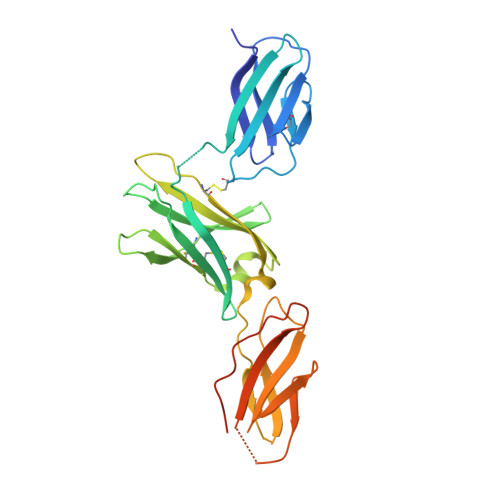
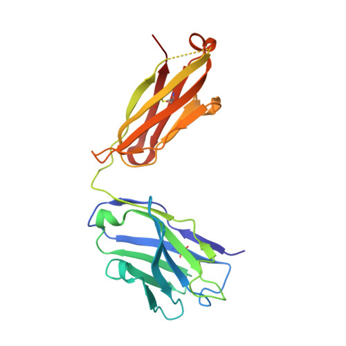
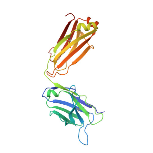
Dual mechanism of interleukin-3 receptor blockade by an anti-cancer antibody

Broughton, S.E.Hercus, T.R.Hardy, M.P.McClure, B.J.Nero, T.L.Dottore, M.Huynh, H.Braley, H.Barry, E.F.Kan, W.L.Dhagat, U.Scotney, P.Hartman, D.Busfield, S.J.Owczarek, C.M.Nash, A.D.Wilson, N.J.Parker, M.W.Lopez, A.F.

(2014) Cell Rep 8: 410-419

  • DOI: https://doi.org/10.1016/j.celrep.2014.06.038
  • Primary Citation Related Structures: 
    4JZJ

  • PubMed Abstract: 

    Interleukin-3 (IL-3) is an activated T cell product that bridges innate and adaptive immunity and contributes to several immunopathologies. Here, we report the crystal structure of the IL-3 receptor α chain (IL3Rα) in complex with the anti-leukemia antibody CSL362 that reveals the N-terminal domain (NTD), a domain also present in the granulocyte-macrophage colony-stimulating factor (GM-CSF), IL-5, and IL-13 receptors, adopting unique "open" and classical "closed" conformations. Although extensive mutational analyses of the NTD epitope of CSL362 show minor overlap with the IL-3 binding site, CSL362 only inhibits IL-3 binding to the closed conformation, indicating alternative mechanisms for blocking IL-3 signaling. Significantly, whereas "open-like" IL3Rα mutants can simultaneously bind IL-3 and CSL362, CSL362 still prevents the assembly of a higher-order IL-3 receptor-signaling complex. The discovery of open forms of cytokine receptors provides the framework for development of potent antibodies that can achieve a "double hit" cytokine receptor blockade.


  • Organizational Affiliation
    • Australian Cancer Research Foundation Rational Drug Discovery Centre, St. Vincent's Institute of Medical Research, Fitzroy, VIC 3065, Australia.

Macromolecule Content 

  • Total Structure Weight: 165.53 kDa 
  • Atom Count: 10,972 
  • Modeled Residue Count: 1,368 
  • Deposited Residue Count: 1,456 
  • Unique protein chains: 3

Macromolecules

Find similar proteins by:|  3D Structure
Entity ID: 1
MoleculeChains  Sequence LengthOrganismDetailsImage
Interleukin-3 receptor subunit alphaA [auth C],
B [auth D]
287Homo sapiensMutation(s): 1 
Gene Names: IL3RA
UniProt & NIH Common Fund Data Resources
Find proteins for P26951 (Homo sapiens)
Explore P26951 
Go to UniProtKB:  P26951
PHAROS:  P26951
GTEx:  ENSG00000185291 
Entity Groups
Sequence Clusters30% Identity50% Identity70% Identity90% Identity95% Identity100% Identity
UniProt GroupP26951
Glycosylation
Glycosylation Sites: 2Go to GlyGen: P26951-1
Sequence Annotations
Expand
Reference Sequence
Find similar proteins by:|  3D Structure
Entity ID: 2
MoleculeChains  Sequence LengthOrganismDetailsImage
Fab Heavy ChainC [auth A],
E [auth H]
221Mus musculusMutation(s): 0 
Find similar proteins by:|  3D Structure
Entity ID: 3
MoleculeChains  Sequence LengthOrganismDetailsImage
Fab Light ChainD [auth B],
F [auth L]
220Mus musculusMutation(s): 0 

Oligosaccharides

Help  
Entity ID: 4
MoleculeChains Length2D Diagram GlycosylationD Interactions
alpha-D-mannopyranose-(1-3)-[alpha-D-mannopyranose-(1-6)]beta-D-mannopyranose-(1-4)-2-acetamido-2-deoxy-beta-D-glucopyranose-(1-4)-[beta-L-fucopyranose-(1-3)][beta-L-fucopyranose-(1-6)]2-acetamido-2-deoxy-beta-D-glucopyranoseG [auth E]7N-Glycosylation
Glycosylation Resources
GlyTouCan: G23711JB
GlyCosmos: G23711JB
GlyGen: G23711JB
Entity ID: 5
MoleculeChains Length2D Diagram GlycosylationD Interactions
beta-D-mannopyranose-(1-4)-2-acetamido-2-deoxy-beta-D-glucopyranose-(1-4)-[beta-L-fucopyranose-(1-3)][beta-L-fucopyranose-(1-6)]2-acetamido-2-deoxy-beta-D-glucopyranoseH [auth F]5N-Glycosylation
Glycosylation Resources
GlyTouCan: G17345YW
GlyCosmos: G17345YW
GlyGen: G17345YW
Entity ID: 6
MoleculeChains Length2D Diagram GlycosylationD Interactions
alpha-L-fucopyranose-(1-3)-[beta-L-fucopyranose-(1-6)]2-acetamido-2-deoxy-beta-D-glucopyranoseI [auth G]3N-Glycosylation
Glycosylation Resources
GlyTouCan: G08300EX
GlyCosmos: G08300EX
GlyGen: G08300EX

Experimental Data & Validation

Experimental Data

  • Method: X-RAY DIFFRACTION
  • Resolution: 2.80 Å
  • R-Value Free:  0.244 (Depositor), 0.249 (DCC) 
  • R-Value Work:  0.185 (Depositor) 
  • R-Value Observed: 0.188 (Depositor) 
Space Group: P 1 21 1
Unit Cell:
Length ( Å )Angle ( ˚ )
a = 88.3α = 90
b = 120.648β = 97.45
c = 92.968γ = 90
Software Package:
Software NamePurpose
PHASERphasing
PHENIXrefinement
PDB_EXTRACTdata extraction
HKL-2000data collection
DENZOdata reduction
SCALEPACKdata scaling
SCALAdata scaling

Structure Validation

View Full Validation Report


Ligand Structure Quality Assessment 


Entry History 

Deposition Data

Revision History  (Full details and data files)

  • Version 1.0: 2014-04-09
    Type: Initial release
  • Version 1.1: 2016-03-16
    Changes: Database references
  • Version 1.2: 2017-11-15
    Changes: Advisory, Refinement description
  • Version 2.0: 2020-07-29
    Type: Remediation
    Reason: Carbohydrate remediation
    Changes: Advisory, Atomic model, Data collection, Database references, Derived calculations, Structure summary
  • Version 2.1: 2024-10-16
    Changes: Data collection, Database references, Structure summary