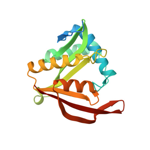
Crystal structure of Ribosomal-protein-alanine N-acetyltransferase from Brucella melitensis in complex with Acetyl CoA
Seattle Structural Genomics Center for Infectious Disease (SSGCID), Abendroth, J., Arakaki, T., Lorimer, D., Edwards, T.E.To be published.