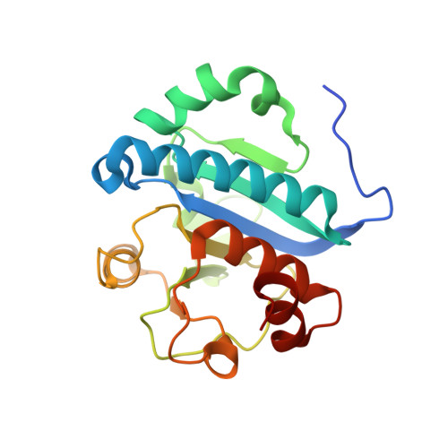
4JWJ

Crystal structure of scTrm10(84)-SAH complex


Experimental Data Snapshot

  • Method: X-RAY DIFFRACTION
  • Resolution: 1.76 Å
  • R-Value Free: 0.251 
  • R-Value Work: 0.218 
  • R-Value Observed: 0.220 

Starting Model: experimental
View more details

wwPDB Validation   3D Report Full Report


Ligand Structure Quality Assessment 


This is version 1.3 of the entry. See complete history


Literature

Crystal structure of tRNA m1G9 methyltransferase Trm10: insight into the catalytic mechanism and recognition of tRNA substrate.

Shao, Z.Yan, W.Peng, J.Zuo, X.Zou, Y.Li, F.Gong, D.Ma, R.Wu, J.Shi, Y.Zhang, Z.Teng, M.Li, X.Gong, Q.

(2014) Nucleic Acids Res 42: 509-525

  • DOI: https://doi.org/10.1093/nar/gkt869
  • Primary Citation of Related Structures:  
    4JWF, 4JWG, 4JWH, 4JWJ

  • PubMed Abstract: 

    Transfer RNA (tRNA) methylation is necessary for the proper biological function of tRNA. The N(1) methylation of guanine at Position 9 (m(1)G9) of tRNA, which is widely identified in eukaryotes and archaea, was found to be catalyzed by the Trm10 family of methyltransferases (MTases). Here, we report the first crystal structures of the tRNA MTase spTrm10 from Schizosaccharomyces pombe in the presence and absence of its methyl donor product S-adenosyl-homocysteine (SAH) and its ortholog scTrm10 from Saccharomyces cerevisiae in complex with SAH. Our crystal structures indicated that the MTase domain (the catalytic domain) of the Trm10 family displays a typical SpoU-TrmD (SPOUT) fold. Furthermore, small angle X-ray scattering analysis reveals that Trm10 behaves as a monomer in solution, whereas other members of the SPOUT superfamily all function as homodimers. We also performed tRNA MTase assays and isothermal titration calorimetry experiments to investigate the catalytic mechanism of Trm10 in vitro. In combination with mutational analysis and electrophoretic mobility shift assays, our results provide insights into the substrate tRNA recognition mechanism of Trm10 family MTases.


  • Organizational Affiliation

    Hefei National Laboratory for Physical Sciences at the Microscale and School of Life Sciences, University of Science and Technology of China, Hefei, Anhui, 230026, People's Republic of China and X-ray Science Division, Advanced Photon Source, Argonne National Laboratory, Argonne, IL 60349, USA.


Macromolecules
Find similar proteins by:  (by identity cutoff)  |  3D Structure
Entity ID: 1
MoleculeChains Sequence LengthOrganismDetailsImage
tRNA (guanine(9)-N1)-methyltransferase
A, B
202Saccharomyces cerevisiae S288CMutation(s): 0 
Gene Names: O0926TRM10YOL093W
EC: 2.1.1.221
UniProt
Find proteins for Q12400 (Saccharomyces cerevisiae (strain ATCC 204508 / S288c))
Explore Q12400 
Go to UniProtKB:  Q12400
Entity Groups  
Sequence Clusters30% Identity50% Identity70% Identity90% Identity95% Identity100% Identity
UniProt GroupQ12400
Sequence Annotations
Expand
  • Reference Sequence
Small Molecules
Ligands 3 Unique
IDChains Name / Formula / InChI Key2D Diagram3D Interactions
SAH
Query on SAH

Download Ideal Coordinates CCD File 
D [auth A],
H [auth B]
S-ADENOSYL-L-HOMOCYSTEINE
C14 H20 N6 O5 S
ZJUKTBDSGOFHSH-WFMPWKQPSA-N
TRS
Query on TRS

Download Ideal Coordinates CCD File 
E [auth A],
J [auth B],
K [auth B]
2-AMINO-2-HYDROXYMETHYL-PROPANE-1,3-DIOL
C4 H12 N O3
LENZDBCJOHFCAS-UHFFFAOYSA-O
SO4
Query on SO4

Download Ideal Coordinates CCD File 
C [auth A],
F [auth A],
G [auth A],
I [auth B]
SULFATE ION
O4 S
QAOWNCQODCNURD-UHFFFAOYSA-L
Experimental Data & Validation

Experimental Data

  • Method: X-RAY DIFFRACTION
  • Resolution: 1.76 Å
  • R-Value Free: 0.251 
  • R-Value Work: 0.218 
  • R-Value Observed: 0.220 
  • Space Group: C 1 2 1
Unit Cell:
Length ( Å )Angle ( ˚ )
a = 131.583α = 90
b = 58.861β = 131.13
c = 99.767γ = 90
Software Package:
Software NamePurpose
DENZOdata reduction
SCALEPACKdata scaling
PHASERphasing
REFMACrefinement
PDB_EXTRACTdata extraction
ADSCdata collection

Structure Validation

View Full Validation Report


Ligand Structure Quality Assessment 


Entry History 

Deposition Data

Revision History  (Full details and data files)

  • Version 1.0: 2013-10-16
    Type: Initial release
  • Version 1.1: 2014-02-12
    Changes: Database references
  • Version 1.2: 2017-11-15
    Changes: Advisory, Refinement description
  • Version 1.3: 2023-11-08
    Changes: Advisory, Data collection, Database references, Derived calculations, Refinement description