4JSD

The X-ray crystal structure of a thermophilic cellobiose binding protein bound with laminaribiose


Experimental Data Snapshot

  • Method: X-RAY DIFFRACTION
  • Resolution: 2.05 Å
  • R-Value Free: 0.203 
  • R-Value Work: 0.184 
  • R-Value Observed: 0.185 

wwPDB Validation   3D Report Full Report


This is version 2.1 of the entry. See complete history


Literature

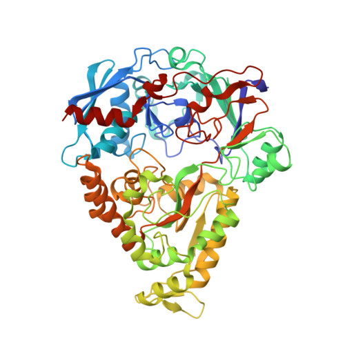
Molecular details of ligand selectivity determinants in a promiscuous beta-glucan periplasmic binding protein.

Munshi, P.Stanley, C.B.Ghimire-Rijal, S.Lu, X.Myles, D.A.Cuneo, M.J.

(2013) BMC Struct Biol 13: 18-18

  • DOI: https://doi.org/10.1186/1472-6807-13-18
  • Primary Citation of Related Structures:  
    4JSD, 4JSO

  • PubMed Abstract: 

    Members of the periplasmic binding protein (PBP) superfamily utilize a highly conserved inter-domain ligand binding site that adapts to specifically bind a chemically diverse range of ligands. This paradigm of PBP ligand binding specificity was recently altered when the structure of the Thermotoga maritima cellobiose-binding protein (tmCBP) was solved. The tmCBP binding site is bipartite, comprising a canonical solvent-excluded region (subsite one), adjacent to a solvent-filled cavity (subsite two) where specific and semi-specific ligand recognition occur, respectively. A molecular level understanding of binding pocket adaptation mechanisms that simultaneously allow both ligand specificity at subsite one and promiscuity at subsite two has potentially important implications in ligand binding and drug design studies. We sought to investigate the determinants of ligand binding selectivity in tmCBP through biophysical characterization of tmCBP in the presence of varying β-glucan oligosaccharides. Crystal structures show that whilst the amino acids that comprise both the tmCBP subsite one and subsite two binding sites remain fixed in conformation regardless of which ligands are present, the rich hydrogen bonding potential of water molecules may facilitate the ordering and the plasticity of this unique PBP binding site. The identification of the roles these water molecules play in ligand recognition suggests potential mechanisms that can be utilized to adapt a single ligand binding site to recognize multiple distinct ligands.


  • Organizational Affiliation

    Neutron Sciences Directorate, Oak Ridge National Laboratory, Oak Ridge, TN 37831, USA. cuneomj@ornl.gov.


Macromolecules
Find similar proteins by:  (by identity cutoff)  |  3D Structure
Entity ID: 1
MoleculeChains Sequence LengthOrganismDetailsImage
Oligopeptide ABC transporter, periplasmic oligopeptide-binding protein582Thermotoga maritima MSB8Mutation(s): 0 
Gene Names: TM0031TM_0031
UniProt
Find proteins for Q9WXN8 (Thermotoga maritima (strain ATCC 43589 / DSM 3109 / JCM 10099 / NBRC 100826 / MSB8))
Explore Q9WXN8 
Go to UniProtKB:  Q9WXN8
Entity Groups  
Sequence Clusters30% Identity50% Identity70% Identity90% Identity95% Identity100% Identity
UniProt GroupQ9WXN8
Sequence Annotations
Expand
  • Reference Sequence
Oligosaccharides

Help

Entity ID: 2
MoleculeChains Length2D Diagram Glycosylation3D Interactions
beta-D-glucopyranose-(1-3)-beta-D-glucopyranose
B
2N/A
Glycosylation Resources
GlyTouCan:  G36535HU
GlyCosmos:  G36535HU
Small Molecules
Ligands 1 Unique
IDChains Name / Formula / InChI Key2D Diagram3D Interactions
CA
Query on CA

Download Ideal Coordinates CCD File 
C [auth A]CALCIUM ION
Ca
BHPQYMZQTOCNFJ-UHFFFAOYSA-N
Biologically Interesting Molecules (External Reference) 1 Unique
Experimental Data & Validation

Experimental Data

  • Method: X-RAY DIFFRACTION
  • Resolution: 2.05 Å
  • R-Value Free: 0.203 
  • R-Value Work: 0.184 
  • R-Value Observed: 0.185 
  • Space Group: P 21 21 21
Unit Cell:
Length ( Å )Angle ( ˚ )
a = 56.641α = 90
b = 89.596β = 90
c = 108.245γ = 90
Software Package:
Software NamePurpose
HKL-3000data collection
PHASERphasing
PHENIXrefinement
HKL-3000data reduction
HKL-3000data scaling

Structure Validation

View Full Validation Report


Entry History 

Deposition Data

Revision History  (Full details and data files)

  • Version 1.0: 2013-10-09
    Type: Initial release
  • Version 1.1: 2014-03-12
    Changes: Database references
  • Version 1.2: 2018-03-07
    Changes: Data collection
  • Version 2.0: 2020-07-29
    Type: Remediation
    Reason: Carbohydrate remediation
    Changes: Atomic model, Data collection, Derived calculations, Structure summary
  • Version 2.1: 2023-09-20
    Changes: Data collection, Database references, Refinement description, Structure summary