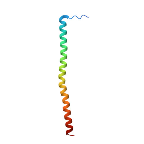
4JOL | pdb_00004jol

Complex structure of AML1-ETO NHR2 domain with HEB fragment


Experimental Data Snapshot

  • Method: X-RAY DIFFRACTION
  • Resolution: 2.91 Å
  • R-Value Free: 
    0.252 (Depositor), 0.252 (DCC) 
  • R-Value Work: 
    0.207 (Depositor), 0.205 (DCC) 
  • R-Value Observed: 
    0.211 (Depositor) 

Starting Model: experimental
View more details

wwPDB Validation 3D Report Full Report

Validation slider image for 4JOL

This is version 1.4 of the entry. See complete history. 

Literature

A stable transcription factor complex nucleated by oligomeric AML1-ETO controls leukaemogenesis.

Sun, X.J., Wang, Z., Wang, L., Jiang, Y., Kost, N., Soong, T.D., Chen, W.Y., Tang, Z., Nakadai, T., Elemento, O., Fischle, W., Melnick, A., Patel, D.J., Nimer, S.D., Roeder, R.G.

(2013) Nature 500: 93-97

  • DOI: https://doi.org/10.1038/nature12287
  • Primary Citation Related Structures: 
    4JOL

  • PubMed Abstract: 

    Transcription factors are frequently altered in leukaemia through chromosomal translocation, mutation or aberrant expression. AML1-ETO, a fusion protein generated by the t(8;21) translocation in acute myeloid leukaemia, is a transcription factor implicated in both gene repression and activation. AML1-ETO oligomerization, mediated by the NHR2 domain, is critical for leukaemogenesis, making it important to identify co-regulatory factors that 'read' the NHR2 oligomerization and contribute to leukaemogenesis. Here we show that, in human leukaemic cells, AML1-ETO resides in and functions through a stable AML1-ETO-containing transcription factor complex (AETFC) that contains several haematopoietic transcription (co)factors. These AETFC components stabilize the complex through multivalent interactions, provide multiple DNA-binding domains for diverse target genes, co-localize genome wide, cooperatively regulate gene expression, and contribute to leukaemogenesis. Within the AETFC complex, AML1-ETO oligomerization is required for a specific interaction between the oligomerized NHR2 domain and a novel NHR2-binding (N2B) motif in E proteins. Crystallographic analysis of the NHR2-N2B complex reveals a unique interaction pattern in which an N2B peptide makes direct contact with side chains of two NHR2 domains as a dimer, providing a novel model of how dimeric/oligomeric transcription factors create a new protein-binding interface through dimerization/oligomerization. Intriguingly, disruption of this interaction by point mutations abrogates AML1-ETO-induced haematopoietic stem/progenitor cell self-renewal and leukaemogenesis. These results reveal new mechanisms of action of AML1-ETO, and provide a potential therapeutic target in t(8;21)-positive acute myeloid leukaemia.


  • Organizational Affiliation: 
    • Laboratory of Biochemistry and Molecular Biology, The Rockefeller University, New York, New York 10065, USA.

Macromolecule Content 

  • Total Structure Weight: 42.63 kDa 
  • Atom Count: 2,425 
  • Modeled Residue Count: 285 
  • Deposited Residue Count: 356 
  • Unique protein chains: 2

Macromolecules

Find similar proteins by:|  3D Structure
Entity ID: 1
MoleculeChains  Sequence LengthOrganismDetailsImage
Protein CBFA2T1
A, B, C, D
64Homo sapiensMutation(s): 0 
Gene Names: RUNX1T1, AML1T1, CBFA2T1, CDR, ETO, MTG8, ZMYND2
UniProt & NIH Common Fund Data Resources
Find proteins for Q06455 (Homo sapiens)
Explore Q06455 
Go to UniProtKB:  Q06455
PHAROS:  Q06455
GTEx:  ENSG00000079102 
Entity Groups
Sequence Clusters30% Identity50% Identity70% Identity90% Identity95% Identity100% Identity
UniProt GroupQ06455
Sequence Annotations
Expand
Reference Sequence
Find similar proteins by:  Sequence   |   3D Structure  
Entity ID: 2
MoleculeChains  Sequence LengthOrganismDetailsImage
Transcription factor 12
E, F, G, H
25Homo sapiensMutation(s): 0 
Gene Names: TCF12, BHLHB20, HEB, HTF4
UniProt & NIH Common Fund Data Resources
Find proteins for Q99081 (Homo sapiens)
Explore Q99081 
Go to UniProtKB:  Q99081
PHAROS:  Q99081
GTEx:  ENSG00000140262 
Entity Groups
Sequence Clusters30% Identity50% Identity70% Identity90% Identity95% Identity100% Identity
UniProt GroupQ99081
Sequence Annotations
Expand
Reference Sequence

Experimental Data & Validation

Experimental Data

  • Method: X-RAY DIFFRACTION
  • Resolution: 2.91 Å
  • R-Value Free:  0.252 (Depositor), 0.252 (DCC) 
  • R-Value Work:  0.207 (Depositor), 0.205 (DCC) 
  • R-Value Observed: 0.211 (Depositor) 
Space Group: P 32
Unit Cell:
Length ( Å )Angle ( ˚ )
a = 139.485α = 90
b = 139.485β = 90
c = 43.169γ = 120
Software Package:
Software NamePurpose
MAR345dtbdata collection
PHENIXmodel building
PHENIXrefinement
HKL-2000data reduction
HKL-2000data scaling
PHENIXphasing

Structure Validation

View Full Validation Report


Entry History 

Deposition Data

Revision History  (Full details and data files)

  • Version 1.0: 2013-06-26
    Type: Initial release
  • Version 1.1: 2013-07-24
    Changes: Database references
  • Version 1.2: 2013-08-07
    Changes: Database references
  • Version 1.3: 2013-08-28
    Changes: Database references
  • Version 1.4: 2023-09-20
    Changes: Data collection, Database references, Refinement description