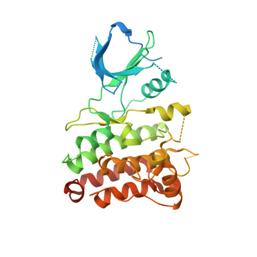
4JOA

Crystal Structure of Human Anaplastic Lymphoma Kinase in complex with 7-azaindole based inhibitor


Experimental Data Snapshot

  • Method: X-RAY DIFFRACTION
  • Resolution: 2.70 Å
  • R-Value Free: 0.290 
  • R-Value Work: 0.251 
  • R-Value Observed: 0.253 

Starting Model: experimental
View more details

wwPDB Validation   3D Report Full Report


Ligand Structure Quality Assessment 


This is version 1.2 of the entry. See complete history


Literature

Discovery of 7-azaindole based anaplastic lymphoma kinase (ALK) inhibitors: wild type and mutant (L1196M) active compounds with unique binding mode

Gummadi, V.R.Rajagopalan, S.Looi, C.Y.Paydar, M.Renukappa, G.A.Ainan, B.R.Krishnamurthy, N.R.Panigrahi, S.K.Mahasweta, K.Raghuramachandran, S.Rajappa, M.Ramanathan, A.Lakshminarasimhan, A.Ramachandra, M.Wong, P.F.Mustafa, M.R.Nanduri, S.Hosahalli, S.

(2013) Bioorg Med Chem Lett 23: 4911-4918

  • DOI: https://doi.org/10.1016/j.bmcl.2013.06.071
  • Primary Citation of Related Structures:  
    4JOA

  • PubMed Abstract: 

    We have identified a novel 7-azaindole series of anaplastic lymphoma kinase (ALK) inhibitors. Compounds 7b, 7 m and 7 n demonstrate excellent potencies in biochemical and cellular assays. X-ray crystal structure of one of the compounds (7 k) revealed a unique binding mode with the benzyl group occupying the back pocket, explaining its potency towards ALK and selectivity over tested kinases particularly Aurora-A. This binding mode is in contrast to that of known ALK inhibitors such as Crizotinib and NVP-TAE684 which occupy the ribose binding pocket, close to DFG motif.


  • Organizational Affiliation

    Aurigene Discovery Technologies Ltd, #39/40, KIADB Industrial Area, Hosur Road, Electronic City Phase-II, Bangalore 560100, India.


Macromolecules
Find similar proteins by:  (by identity cutoff)  |  3D Structure
Entity ID: 1
MoleculeChains Sequence LengthOrganismDetailsImage
ALK tyrosine kinase receptor344Homo sapiensMutation(s): 1 
Gene Names: ALK
EC: 2.7.10.1
UniProt & NIH Common Fund Data Resources
Find proteins for Q9UM73 (Homo sapiens)
Explore Q9UM73 
Go to UniProtKB:  Q9UM73
PHAROS:  Q9UM73
GTEx:  ENSG00000171094 
Entity Groups  
Sequence Clusters30% Identity50% Identity70% Identity90% Identity95% Identity100% Identity
UniProt GroupQ9UM73
Sequence Annotations
Expand
  • Reference Sequence
Small Molecules
Ligands 1 Unique
IDChains Name / Formula / InChI Key2D Diagram3D Interactions
3DK
Query on 3DK

Download Ideal Coordinates CCD File 
B [auth A]3-[1-(2,5-difluorobenzyl)-1H-pyrazol-4-yl]-5-(1-methyl-1H-pyrazol-4-yl)-1H-pyrrolo[2,3-b]pyridine
C21 H16 F2 N6
SDWMCZUOCJDBOJ-UHFFFAOYSA-N
Binding Affinity Annotations 
IDSourceBinding Affinity
3DK PDBBind:  4JOA IC50: 29 (nM) from 1 assay(s)
BindingDB:  4JOA IC50: min: 29, max: 1592 (nM) from 3 assay(s)
Experimental Data & Validation

Experimental Data

  • Method: X-RAY DIFFRACTION
  • Resolution: 2.70 Å
  • R-Value Free: 0.290 
  • R-Value Work: 0.251 
  • R-Value Observed: 0.253 
  • Space Group: P 21 21 21
Unit Cell:
Length ( Å )Angle ( ˚ )
a = 51.794α = 90
b = 56.503β = 90
c = 104.536γ = 90
Software Package:
Software NamePurpose
CrystalCleardata collection
MOLREPphasing
REFMACrefinement
DENZOdata reduction
SCALEPACKdata scaling

Structure Validation

View Full Validation Report


Ligand Structure Quality Assessment 


Entry History 

Deposition Data

Revision History  (Full details and data files)

  • Version 1.0: 2013-07-17
    Type: Initial release
  • Version 1.1: 2014-04-23
    Changes: Database references
  • Version 1.2: 2023-11-08
    Changes: Data collection, Database references, Derived calculations, Refinement description