4JNA | pdb_00004jna

Crystal structure of the DepH complex with dimethyl-FK228


Experimental Data Snapshot

  • Method: X-RAY DIFFRACTION
  • Resolution: 2.00 Å
  • R-Value Free: 
    0.222 (Depositor), 0.224 (DCC) 
  • R-Value Work: 
    0.152 (Depositor), 0.155 (DCC) 
  • R-Value Observed: 
    0.155 (Depositor) 

Starting Model: experimental
View more details

wwPDB Validation 3D Report Full Report

Validation slider image for 4JNA

Ligand Structure Quality Assessment 


This is version 2.0 of the entry. See complete history

Literature

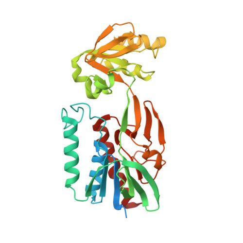
The structural basis of an NADP+-independent dithiol oxidase in FK228 biosynthesis.

Li, J.Wang, C.Zhang, Z.M.Cheng, Y.Q.Zhou, J.

(2014) Sci Rep 4: 4145-4145

  • DOI: https://doi.org/10.1038/srep04145
  • Primary Citation Related Structures: 
    4JN9, 4JNA

  • PubMed Abstract: 

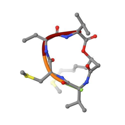
    The disulfide bond is unusual in natural products and critical for thermal stability, cell permeability and bioactivity. DepH from Chromobacterium violaceum No. 968 is an FAD-dependent enzyme responsible for catalyzing the disulfide bond formation of FK228, an anticancer prodrug approved for the treatment of cutaneous T-cell lymphoma. Here we report the crystal structures of DepH and DepH complexed with a substrate analogue S,S'-dimethyl FK228 at 1.82 Å and 2.00 Å, respectively. Structural and biochemical analyses revealed that DepH, in contrast to the well characterized low molecular weight thioredoxin reductases (LMW TrxRs), is an NADP(+)-independent dithiol oxidase. DepH not only lacks a conserved GGGDXAXE motif necessary for NADP(+) binding in the canonical LMW TrxRs, but also contains a 11-residue sequence which physically impedes the binding of NADP(+). These observations explain the difference between NADP(+)-independent small molecule dithiol oxidases and NADP(+)-dependent thioredoxin reductases and provide insights for understanding the catalytic mechanism of dithiol oxidases involved in natural product biosynthesis.


  • Organizational Affiliation
    • 1] State Key Laboratory of Bio-organic and Natural Products Chemistry, Shanghai Institute of Organic Chemistry, Chinese Academy of Sciences, Shanghai 200032, China [2].

Macromolecule Content 

  • Total Structure Weight: 76.76 kDa 
  • Atom Count: 5,012 
  • Modeled Residue Count: 599 
  • Deposited Residue Count: 690 
  • Unique protein chains: 2

Macromolecules

Find similar proteins by:|  3D Structure
Entity ID: 1
MoleculeChains  Sequence LengthOrganismDetailsImage
DepH
A, B
340Chromobacterium violaceumMutation(s): 0 
Gene Names: depH
UniProt
Find proteins for A4ZPY8 (Chromobacterium violaceum)
Explore A4ZPY8 
Go to UniProtKB:  A4ZPY8
Entity Groups
Sequence Clusters30% Identity50% Identity70% Identity90% Identity95% Identity100% Identity
UniProt GroupA4ZPY8
Sequence Annotations
Expand
Reference Sequence
Find similar proteins by:  Sequence   |   3D Structure  
Entity ID: 2
MoleculeChains  Sequence LengthOrganismDetailsImage
Dimethyl FK228C [auth H],
D [auth I]
5N/AMutation(s): 0 

Small Molecules

Ligands 5 Unique
IDChains Name / Formula / InChI Key2D Diagram3D Interactions
FAD

Query on FAD


Download:Ideal Coordinates CCD File
E [auth A],
J [auth B]
FLAVIN-ADENINE DINUCLEOTIDE
C27 H33 N9 O15 P2
VWWQXMAJTJZDQX-UYBVJOGSSA-N
PO4

Query on PO4


Download:Ideal Coordinates CCD File
I [auth A]PHOSPHATE ION
O4 P
NBIIXXVUZAFLBC-UHFFFAOYSA-K
GOL

Query on GOL


Download:Ideal Coordinates CCD File
N [auth B]GLYCEROL
C3 H8 O3
PEDCQBHIVMGVHV-UHFFFAOYSA-N
EDO

Query on EDO


Download:Ideal Coordinates CCD File
G [auth A],
H [auth A],
L [auth B],
M [auth B]
1,2-ETHANEDIOL
C2 H6 O2
LYCAIKOWRPUZTN-UHFFFAOYSA-N
CL

Query on CL


Download:Ideal Coordinates CCD File
F [auth A],
K [auth B]
CHLORIDE ION
Cl
VEXZGXHMUGYJMC-UHFFFAOYSA-M
Modified Residues  1 Unique
IDChains TypeFormula2D DiagramParent
060
Query on 060
C [auth H],
D [auth I]
D-PEPTIDE LINKINGC4 H9 N O2 S

--

DBU
Query on DBU
C [auth H],
D [auth I]
PEPTIDE LINKINGC4 H7 N O2THR
DVA
Query on DVA
C [auth H],
D [auth I]
D-PEPTIDE LINKINGC5 H11 N O2

--

Biologically Interesting Molecules (External Reference) 

1 Unique

Experimental Data & Validation

Experimental Data

  • Method: X-RAY DIFFRACTION
  • Resolution: 2.00 Å
  • R-Value Free:  0.222 (Depositor), 0.224 (DCC) 
  • R-Value Work:  0.152 (Depositor), 0.155 (DCC) 
  • R-Value Observed: 0.155 (Depositor) 
Space Group: P 43 21 2
Unit Cell:
Length ( Å )Angle ( ˚ )
a = 154.283α = 90
b = 154.283β = 90
c = 71.828γ = 90
Software Package:
Software NamePurpose
DENZOdata reduction
SCALEPACKdata scaling
PHASERphasing
PHENIXrefinement
PDB_EXTRACTdata extraction
HKL-2000data collection

Structure Validation

View Full Validation Report


Ligand Structure Quality Assessment 


Entry History 

Deposition Data

Revision History  (Full details and data files)

  • Version 1.0: 2014-03-05
    Type: Initial release
  • Version 1.1: 2014-04-09
    Changes: Database references
  • Version 1.2: 2017-11-15
    Changes: Advisory, Refinement description
  • Version 1.3: 2023-09-20
    Changes: Data collection, Database references, Derived calculations, Refinement description
  • Version 2.0: 2023-11-15
    Changes: Atomic model, Data collection, Derived calculations