


Docking to a water-filled model binding site in Cytochrome c Peroxidase
Barelier, S., Boyce, S.E., Fish, I., Fischer, M., Goodin, D.B., Shoichet, B.K.To be published.
Experimental Data Snapshot
Entity ID: 1 | |||||
|---|---|---|---|---|---|
| Molecule | Chains | Sequence Length | Organism | Details | Image |
| Cytochrome c peroxidase | 289 | Saccharomyces cerevisiae RM11-1a | Mutation(s): 2 Gene Names: CCP1 CCP CPO YKR066C, SCRG_04081 EC: 1.11.1.5 (PDB Primary Data), 1.11.1 (UniProt) | 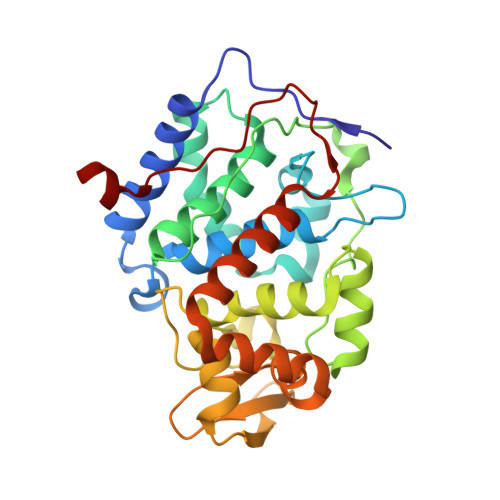 | |
UniProt | |||||
Entity Groups | |||||
| Sequence Clusters | 30% Identity50% Identity70% Identity90% Identity95% Identity100% Identity | ||||
| UniProt Group | B3LRE1 | ||||
Sequence AnnotationsExpand | |||||
Reference Sequence | |||||
| Ligands 3 Unique | |||||
|---|---|---|---|---|---|
| ID | Chains | Name / Formula / InChI Key | 2D Diagram | 3D Interactions | |
| HEM Download:Ideal Coordinates CCD File | B [auth A] | PROTOPORPHYRIN IX CONTAINING FE C34 H32 Fe N4 O4 KABFMIBPWCXCRK-RGGAHWMASA-L |  | ||
| 1LW Download:Ideal Coordinates CCD File | C [auth A] | 5,6,7,8-tetrahydrothieno[2,3-b]quinolin-4-amine C11 H12 N2 S PMWMMNQYOBSEDT-UHFFFAOYSA-N |  | ||
| MES Download:Ideal Coordinates CCD File | D [auth A] | 2-(N-MORPHOLINO)-ETHANESULFONIC ACID C6 H13 N O4 S SXGZJKUKBWWHRA-UHFFFAOYSA-N |  | ||
| Length ( Å ) | Angle ( ˚ ) |
|---|---|
| a = 106.82 | α = 90 |
| b = 74.8 | β = 90 |
| c = 50.97 | γ = 90 |
| Software Name | Purpose |
|---|---|
| PHENIX | refinement |
| PDB_EXTRACT | data extraction |
| ADSC | data collection |
| XDS | data reduction |
| XDS | data scaling |


