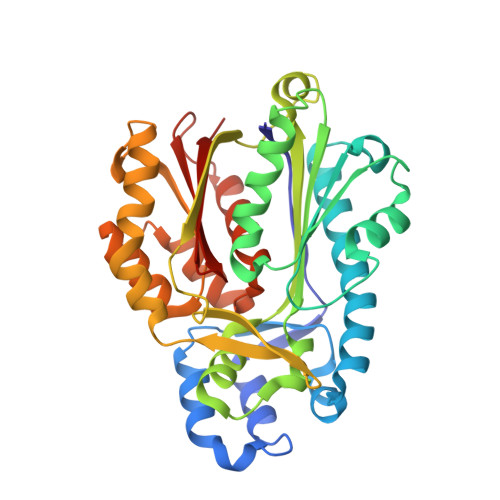
Crystal structure of Mycobacterium tuberculosis polyketide synthase 11 (PKS11) reveals intermediates in the synthesis of methyl-branched alkylpyrones.
Gokulan, K., O'Leary, S.E., Russell, W.K., Russell, D.H., Lalgondar, M., Begley, T.P., Ioerger, T.R., Sacchettini, J.C.(2013) J Biological Chem 288: 16484-16494
- PubMed: 23615910
- DOI: https://doi.org/10.1074/jbc.M113.468892
- Primary Citation of Related Structures:
4JAO, 4JAP, 4JAQ, 4JAR, 4JAT, 4JD3 - PubMed Abstract:
PKS11 is one of three type III polyketide synthases (PKSs) identified in Mycobacterium tuberculosis. Although many PKSs in M. tuberculosis have been implicated in producing complex cell wall glycolipids, the biological function of PKS11 is unknown. PKS11 has previously been proposed to synthesize alkylpyrones from fatty acid substrates. We solved the crystal structure of M. tuberculosis PKS11 and found the overall fold to be similar to other type III PKSs. PKS11 has a deep hydrophobic tunnel proximal to the active site Cys-138 to accommodate substrates. We observed electron density in this tunnel from a co-purified molecule that was identified by mass spectrometry to be palmitate. Co-crystallization with malonyl-CoA (MCoA) or methylmalonyl-CoA (MMCoA) led to partial turnover of the substrate, resulting in trapped intermediates. Reconstitution of the reaction in solution confirmed that both co-factors are required for optimal activity, and kinetic analysis shows that MMCoA is incorporated first, then MCoA, followed by lactonization to produce methyl-branched alkylpyrones.
- Departments of Biochemistry and Biophysics, College Station, Texas 77843.
Organizational Affiliation:


