4JA4 | pdb_00004ja4

Inward open conformation of the xylose transporter XylE from E. coli


Experimental Data Snapshot

  • Method: X-RAY DIFFRACTION
  • Resolution: 4.20 Å
  • R-Value Free: 
    0.318 (Depositor), 0.323 (DCC) 
  • R-Value Work: 
    0.288 (Depositor), 0.299 (DCC) 
  • R-Value Observed: 
    0.290 (Depositor) 

wwPDB Validation 3D Report Full Report

Validation slider image for 4JA4

This is version 1.2 of the entry. See complete history

Literature

Structural basis for substrate transport in the GLUT-homology family of monosaccharide transporters.

Quistgaard, E.M.Low, C.Moberg, P.Tresaugues, L.Nordlund, P.

(2013) Nat Struct Mol Biol 20: 766-768

  • DOI: https://doi.org/10.1038/nsmb.2569
  • Primary Citation Related Structures: 
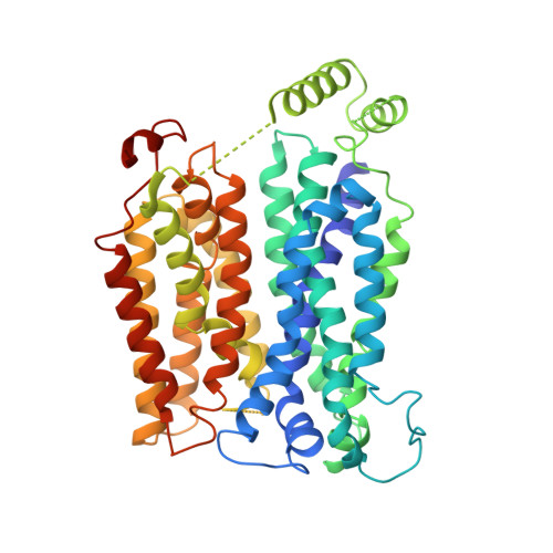
    4JA3, 4JA4

  • PubMed Abstract: 

    Here we present two structures of the major facilitator (MFS) xylose transporter XylE from Escherichia coli in inward open and partially occluded inward open conformations. These structures provide key information about the transport cycle of XylE and the closely related human GLUT transporters. This is, to our knowledge, the first MFS transporter structure determined in more than one conformational state, which may establish XylE as an important MFS model protein.


  • Organizational Affiliation
    • Department of Medical Biochemistry and Biophysics, Karolinska Institutet, Stockholm, Sweden.

Macromolecule Content 

  • Total Structure Weight: 158.89 kDa 
  • Atom Count: 10,130 
  • Modeled Residue Count: 1,317 
  • Deposited Residue Count: 1,455 
  • Unique protein chains: 1

Macromolecules

Find similar proteins by:|  3D Structure
Entity ID: 1
MoleculeChains  Sequence LengthOrganismDetailsImage
D-xylose-proton symporter
A, B, C
485Escherichia coli K-12Mutation(s): 0 
Gene Names: xylEb4031JW3991
Membrane Entity: Yes 
UniProt
Find proteins for P0AGF4 (Escherichia coli (strain K12))
Explore P0AGF4 
Go to UniProtKB:  P0AGF4
Entity Groups
Sequence Clusters30% Identity50% Identity70% Identity90% Identity95% Identity100% Identity
UniProt GroupP0AGF4
Sequence Annotations
Expand
Reference Sequence

Small Molecules

Ligands 1 Unique
IDChains Name / Formula / InChI Key2D Diagram3D Interactions
CD

Query on CD


Download:Ideal Coordinates CCD File
D [auth A]CADMIUM ION
Cd
WLZRMCYVCSSEQC-UHFFFAOYSA-N

Experimental Data & Validation

Experimental Data

  • Method: X-RAY DIFFRACTION
  • Resolution: 4.20 Å
  • R-Value Free:  0.318 (Depositor), 0.323 (DCC) 
  • R-Value Work:  0.288 (Depositor), 0.299 (DCC) 
  • R-Value Observed: 0.290 (Depositor) 
Space Group: P 61
Unit Cell:
Length ( Å )Angle ( ˚ )
a = 125.82α = 90
b = 125.82β = 90
c = 380.68γ = 120
Software Package:
Software NamePurpose
XDSdata scaling
PHASERphasing
PHENIXrefinement
XDSdata reduction
XSCALEdata scaling

Structure Validation

View Full Validation Report


Entry History 

Deposition Data

Revision History  (Full details and data files)

  • Version 1.0: 2013-05-01
    Type: Initial release
  • Version 1.1: 2013-07-17
    Changes: Database references
  • Version 1.2: 2024-02-28
    Changes: Data collection, Database references, Derived calculations