4J5S | pdb_00004j5s

TARG1 (C6orf130), Terminal ADP-ribose Glycohydrolase 1 ADP-ribose complex


Experimental Data Snapshot

  • Method: X-RAY DIFFRACTION
  • Resolution: 1.55 Å
  • R-Value Free: 
    0.175 (Depositor), 0.182 (DCC) 
  • R-Value Work: 
    0.137 (Depositor), 0.147 (DCC) 
  • R-Value Observed: 
    0.139 (Depositor) 

wwPDB Validation 3D Report Full Report

Validation slider image for 4J5S

Ligand Structure Quality Assessment 


This is version 1.3 of the entry. See complete history

Literature

Deficiency of terminal ADP-ribose protein glycohydrolase TARG1/C6orf130 in neurodegenerative disease.

Sharifi, R.Morra, R.Appel, C.D.Tallis, M.Chioza, B.Jankevicius, G.Simpson, M.A.Matic, I.Ozkan, E.Golia, B.Schellenberg, M.J.Weston, R.Williams, J.G.Rossi, M.N.Galehdari, H.Krahn, J.Wan, A.Trembath, R.C.Crosby, A.H.Ahel, D.Hay, R.Ladurner, A.G.Timinszky, G.Williams, R.S.Ahel, I.

(2013) EMBO J 32: 1225-1237

  • DOI: https://doi.org/10.1038/emboj.2013.51
  • Primary Citation Related Structures: 
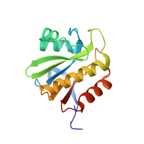
    4J5Q, 4J5R, 4J5S

  • PubMed Abstract: 

    Adenosine diphosphate (ADP)-ribosylation is a post-translational protein modification implicated in the regulation of a range of cellular processes. A family of proteins that catalyse ADP-ribosylation reactions are the poly(ADP-ribose) (PAR) polymerases (PARPs). PARPs covalently attach an ADP-ribose nucleotide to target proteins and some PARP family members can subsequently add additional ADP-ribose units to generate a PAR chain. The hydrolysis of PAR chains is catalysed by PAR glycohydrolase (PARG). PARG is unable to cleave the mono(ADP-ribose) unit directly linked to the protein and although the enzymatic activity that catalyses this reaction has been detected in mammalian cell extracts, the protein(s) responsible remain unknown. Here, we report the homozygous mutation of the c6orf130 gene in patients with severe neurodegeneration, and identify C6orf130 as a PARP-interacting protein that removes mono(ADP-ribosyl)ation on glutamate amino acid residues in PARP-modified proteins. X-ray structures and biochemical analysis of C6orf130 suggest a mechanism of catalytic reversal involving a transient C6orf130 lysyl-(ADP-ribose) intermediate. Furthermore, depletion of C6orf130 protein in cells leads to proliferation and DNA repair defects. Collectively, our data suggest that C6orf130 enzymatic activity has a role in the turnover and recycling of protein ADP-ribosylation, and we have implicated the importance of this protein in supporting normal cellular function in humans.


  • Organizational Affiliation
    • Biomedical Sciences Division, Human Genetics Research Centre, St George's University of London, London, UK. rsharifi@sgul.ac.uk

Macromolecule Content 

  • Total Structure Weight: 69.21 kDa 
  • Atom Count: 5,783 
  • Modeled Residue Count: 567 
  • Deposited Residue Count: 584 
  • Unique protein chains: 1

Macromolecules

Find similar proteins by:|  3D Structure
Entity ID: 1
MoleculeChains  Sequence LengthOrganismDetailsImage
O-acetyl-ADP-ribose deacetylase 1
A, B, C, D
146Homo sapiensMutation(s): 0 
Gene Names: OARD1C6orf130
EC: 3.5.1 (PDB Primary Data), 3.2.2 (UniProt)
UniProt & NIH Common Fund Data Resources
Find proteins for Q9Y530 (Homo sapiens)
Explore Q9Y530 
Go to UniProtKB:  Q9Y530
PHAROS:  Q9Y530
GTEx:  ENSG00000124596 
Entity Groups
Sequence Clusters30% Identity50% Identity70% Identity90% Identity95% Identity100% Identity
UniProt GroupQ9Y530
Sequence Annotations
Expand
Reference Sequence

Small Molecules

Ligands 5 Unique
IDChains Name / Formula / InChI Key2D Diagram3D Interactions
ZZC

Query on ZZC


Download:Ideal Coordinates CCD File
BA [auth D],
E [auth A],
N [auth B],
X [auth C]
[(2R,3S,4R,5R)-5-(6-AMINOPURIN-9-YL)-3,4-DIHYDROXY-OXOLAN-2-YL]METHYL [[(2R,3R)-2,3-DIHYDROXY-4-OXO-PENTOXY]-OXIDO-PHOSPHORYL] PHOSPHATE
C15 H21 N5 O13 P2
IZTPERUWNZCNNH-GHVQHMAVSA-L
SBE

Query on SBE


Download:Ideal Coordinates CCD File
O [auth B]1,3,2-DIOXABOROLAN-2-OL
C2 H5 B O3
ZBEDLGKSWBORBS-UHFFFAOYSA-N
BO4

Query on BO4


Download:Ideal Coordinates CCD File
F [auth A]BORATE ION
B H4 O4
KCFLOKKYWBPKFN-UHFFFAOYSA-N
EDO

Query on EDO


Download:Ideal Coordinates CCD File
AA [auth C]
DA [auth D]
EA [auth D]
FA [auth D]
GA [auth D]
AA [auth C],
DA [auth D],
EA [auth D],
FA [auth D],
GA [auth D],
H [auth A],
I [auth A],
J [auth A],
K [auth A],
L [auth A],
M [auth A],
Q [auth B],
R [auth B],
S [auth B],
T [auth B],
U [auth B],
V [auth B],
W [auth B],
Z [auth C]
1,2-ETHANEDIOL
C2 H6 O2
LYCAIKOWRPUZTN-UHFFFAOYSA-N
CL

Query on CL


Download:Ideal Coordinates CCD File
CA [auth D],
G [auth A],
P [auth B],
Y [auth C]
CHLORIDE ION
Cl
VEXZGXHMUGYJMC-UHFFFAOYSA-M

Experimental Data & Validation

Experimental Data

  • Method: X-RAY DIFFRACTION
  • Resolution: 1.55 Å
  • R-Value Free:  0.175 (Depositor), 0.182 (DCC) 
  • R-Value Work:  0.137 (Depositor), 0.147 (DCC) 
  • R-Value Observed: 0.139 (Depositor) 
Space Group: P 1 21 1
Unit Cell:
Length ( Å )Angle ( ˚ )
a = 73.375α = 90
b = 56.221β = 94.47
c = 73.788γ = 90
Software Package:
Software NamePurpose
SCALEPACKdata scaling
PHASERphasing
PHENIXrefinement
PDB_EXTRACTdata extraction
HKL-2000data collection
DENZOdata reduction

Structure Validation

View Full Validation Report


Ligand Structure Quality Assessment 


Entry History 

Deposition Data

Revision History  (Full details and data files)

  • Version 1.0: 2013-03-27
    Type: Initial release
  • Version 1.1: 2013-05-01
    Changes: Database references
  • Version 1.2: 2013-07-03
    Changes: Database references
  • Version 1.3: 2024-10-09
    Changes: Data collection, Database references, Derived calculations, Structure summary