4J4T | pdb_00004j4t

Crystal Structure of FabI from F. tularensis in complex with novel inhibitors based on the benzimidazole scaffold


Experimental Data Snapshot

  • Method: X-RAY DIFFRACTION
  • Resolution: 2.34 Å
  • R-Value Free: 
    0.241 (Depositor), 0.238 (DCC) 
  • R-Value Work: 
    0.199 (Depositor), 0.197 (DCC) 
  • R-Value Observed: 
    0.201 (Depositor) 

Starting Model: experimental
View more details

wwPDB Validation 3D Report Full Report

Validation slider image for 4J4T

Ligand Structure Quality Assessment 


This is version 1.4 of the entry. See complete history

Literature

Structural and biological evaluation of a novel series of benzimidazole inhibitors of Francisella tularensis enoyl-ACP reductase (FabI).

Mehboob, S.Song, J.Hevener, K.E.Su, P.C.Boci, T.Brubaker, L.Truong, L.Mistry, T.Deng, J.Cook, J.L.Santarsiero, B.D.Ghosh, A.K.Johnson, M.E.

(2015) Bioorg Med Chem Lett 25: 1292-1296

  • DOI: https://doi.org/10.1016/j.bmcl.2015.01.048
  • Primary Citation Related Structures: 
    4J1N, 4J3F, 4J4T

  • PubMed Abstract: 

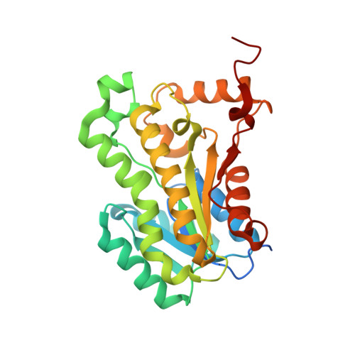
    Francisella tularensis, the causative agent of tularemia, presents a significant biological threat and is a Category A priority pathogen due to its potential for weaponization. The bacterial FASII pathway is a viable target for the development of novel antibacterial agents treating Gram-negative infections. Here we report the advancement of a promising series of benzimidazole FabI (enoyl-ACP reductase) inhibitors to a second-generation using a systematic, structure-guided lead optimization strategy, and the determination of several co-crystal structures that confirm the binding mode of designed inhibitors. These compounds display an improved low nanomolar enzymatic activity as well as promising low microgram/mL antibacterial activity against both F. tularensis and Staphylococcus aureus and its methicillin-resistant strain (MRSA). The improvements in activity accompanying structural modifications lead to a better understanding of the relationship between the chemical structure and biological activity that encompasses both enzymatic and whole-cell activity.


  • Organizational Affiliation
    • Center for Pharmaceutical Biotechnology, University of Illinois at Chicago, Chicago, IL 60607, United States; Novalex Therapeutics, 2242 W. Harrison, Chicago, IL 60612, United States. Electronic address: shahila@uic.edu.

Macromolecule Content 

  • Total Structure Weight: 248.64 kDa 
  • Atom Count: 16,987 
  • Modeled Residue Count: 2,072 
  • Deposited Residue Count: 2,240 
  • Unique protein chains: 1

Macromolecules

Find similar proteins by:|  3D Structure
Entity ID: 1
MoleculeChains  Sequence LengthOrganismDetailsImage
Enoyl-[acyl-carrier-protein] reductase [NADH]
A, B, C, D, E
A, B, C, D, E, F, G, H
280Francisella tularensis subsp. tularensis SCHU S4Mutation(s): 0 
Gene Names: fabIFTT_0782
EC: 1.3.1.9
UniProt
Find proteins for Q5NGQ3 (Francisella tularensis subsp. tularensis (strain SCHU S4 / Schu 4))
Explore Q5NGQ3 
Go to UniProtKB:  Q5NGQ3
Entity Groups
Sequence Clusters30% Identity50% Identity70% Identity90% Identity95% Identity100% Identity
UniProt GroupQ5NGQ3
Sequence Annotations
Expand
Reference Sequence

Small Molecules

Ligands 5 Unique
IDChains Name / Formula / InChI Key2D Diagram3D Interactions
NAD

Query on NAD


Download:Ideal Coordinates CCD File
AA [auth F]
DA [auth G]
FA [auth H]
I [auth A]
M [auth B]
AA [auth F],
DA [auth G],
FA [auth H],
I [auth A],
M [auth B],
R [auth C],
U [auth D],
Y [auth E]
NICOTINAMIDE-ADENINE-DINUCLEOTIDE
C21 H27 N7 O14 P2
BAWFJGJZGIEFAR-NNYOXOHSSA-N
1JU

Query on 1JU


Download:Ideal Coordinates CCD File
BA [auth F]
EA [auth G]
GA [auth H]
J [auth A]
N [auth B]
BA [auth F],
EA [auth G],
GA [auth H],
J [auth A],
N [auth B],
S [auth C],
V [auth D],
Z [auth E]
1-(1,3-benzodioxol-5-ylmethyl)-5,6,7,8-tetrahydro-1H-naphtho[2,3-d]imidazole
C19 H18 N2 O2
GFWJMMLPXBFICR-UHFFFAOYSA-N
GOL

Query on GOL


Download:Ideal Coordinates CCD File
IA [auth H]
JA [auth H]
K [auth A]
L [auth A]
P [auth B]
IA [auth H],
JA [auth H],
K [auth A],
L [auth A],
P [auth B],
T [auth C],
W [auth D]
GLYCEROL
C3 H8 O3
PEDCQBHIVMGVHV-UHFFFAOYSA-N
ACT

Query on ACT


Download:Ideal Coordinates CCD File
Q [auth B],
X [auth D]
ACETATE ION
C2 H3 O2
QTBSBXVTEAMEQO-UHFFFAOYSA-M
NA

Query on NA


Download:Ideal Coordinates CCD File
CA [auth F],
HA [auth H],
O [auth B]
SODIUM ION
Na
FKNQFGJONOIPTF-UHFFFAOYSA-N

Experimental Data & Validation

Experimental Data

  • Method: X-RAY DIFFRACTION
  • Resolution: 2.34 Å
  • R-Value Free:  0.241 (Depositor), 0.238 (DCC) 
  • R-Value Work:  0.199 (Depositor), 0.197 (DCC) 
  • R-Value Observed: 0.201 (Depositor) 
Space Group: P 21 21 21
Unit Cell:
Length ( Å )Angle ( ˚ )
a = 85.398α = 90
b = 123.349β = 90
c = 202.653γ = 90
Software Package:
Software NamePurpose
HKL-2000data collection
PHASERphasing
REFMACrefinement
XDSdata reduction
XDSdata scaling

Structure Validation

View Full Validation Report


Ligand Structure Quality Assessment 


Entry History 

Deposition Data

Revision History  (Full details and data files)

  • Version 1.0: 2014-07-23
    Type: Initial release
  • Version 1.1: 2014-11-12
    Changes: Structure summary
  • Version 1.2: 2015-03-04
    Changes: Database references
  • Version 1.3: 2015-03-18
    Changes: Database references
  • Version 1.4: 2023-09-20
    Changes: Data collection, Database references, Derived calculations, Refinement description