4ISE

Human glucokinase in complex with novel activator (2S)-3-cyclohexyl-2-(6-fluoro-4-oxoquinazolin-3(4H)-yl)-N-(1,3-thiazol-2-yl)propanamide


Experimental Data Snapshot

  • Method: X-RAY DIFFRACTION
  • Resolution: 1.78 Å
  • R-Value Free: 0.227 
  • R-Value Work: 0.199 
  • R-Value Observed: 0.200 

wwPDB Validation   3D Report Full Report


Ligand Structure Quality Assessment 


This is version 1.3 of the entry. See complete history


Literature

Design, synthesis and SAR of novel glucokinase activators.

Cheruvallath, Z.S.Gwaltney, S.L.Sabat, M.Tang, M.Feng, J.Wang, H.Miura, J.Guntupalli, P.Jennings, A.Hosfield, D.Lee, B.Wu, Y.

(2013) Bioorg Med Chem Lett 23: 2166-2171

  • DOI: https://doi.org/10.1016/j.bmcl.2013.01.093
  • Primary Citation of Related Structures:  
    4ISE

  • PubMed Abstract: 

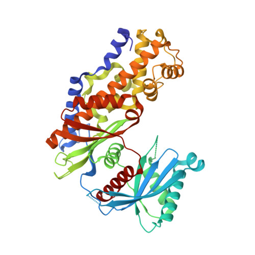
    Guided by co-crystal structures of compounds 15, 22 and 30, an SBDD approach led to the discovery of the 6-methyl pyridone series as a novel class of GKAs that potently activate GK in enzyme and cell assays. Anti-diabetic OGTT efficacy was demonstrated with 54 in a mouse model of type 2 diabetes.


  • Organizational Affiliation

    Takeda California, 10410 Science Center Drive, San Diego, CA 92121, USA.


Macromolecules
Find similar proteins by:  (by identity cutoff)  |  3D Structure
Entity ID: 1
MoleculeChains Sequence LengthOrganismDetailsImage
Glucokinase458Homo sapiensMutation(s): 0 
Gene Names: GCKHXK4 Isoform II
EC: 2.7.1.2
UniProt & NIH Common Fund Data Resources
Find proteins for P35557 (Homo sapiens)
Explore P35557 
Go to UniProtKB:  P35557
PHAROS:  P35557
GTEx:  ENSG00000106633 
Entity Groups  
Sequence Clusters30% Identity50% Identity70% Identity90% Identity95% Identity100% Identity
UniProt GroupP35557
Sequence Annotations
Expand
  • Reference Sequence
Small Molecules
Ligands 3 Unique
IDChains Name / Formula / InChI Key2D Diagram3D Interactions
1FW
Query on 1FW

Download Ideal Coordinates CCD File 
C [auth A](2S)-3-cyclohexyl-2-(6-fluoro-4-oxoquinazolin-3(4H)-yl)-N-(1,3-thiazol-2-yl)propanamide
C20 H21 F N4 O2 S
HETBALZVBPDEQB-KRWDZBQOSA-N
GLC
Query on GLC

Download Ideal Coordinates CCD File 
B [auth A]alpha-D-glucopyranose
C6 H12 O6
WQZGKKKJIJFFOK-DVKNGEFBSA-N
IOD
Query on IOD

Download Ideal Coordinates CCD File 
D [auth A]IODIDE ION
I
XMBWDFGMSWQBCA-UHFFFAOYSA-M
Binding Affinity Annotations 
IDSourceBinding Affinity
1FW BindingDB:  4ISE EC50: 4000 (nM) from 1 assay(s)
Experimental Data & Validation

Experimental Data

  • Method: X-RAY DIFFRACTION
  • Resolution: 1.78 Å
  • R-Value Free: 0.227 
  • R-Value Work: 0.199 
  • R-Value Observed: 0.200 
  • Space Group: P 21 21 21
Unit Cell:
Length ( Å )Angle ( ˚ )
a = 49.483α = 90
b = 77.646β = 90
c = 119.274γ = 90
Software Package:
Software NamePurpose
HKL-2000data collection
MOLREPphasing
REFMACrefinement
HKL-2000data reduction
HKL-2000data scaling

Structure Validation

View Full Validation Report


Ligand Structure Quality Assessment 


Entry History 

Deposition Data

Revision History  (Full details and data files)

  • Version 1.0: 2013-03-20
    Type: Initial release
  • Version 1.1: 2013-04-03
    Changes: Database references
  • Version 1.2: 2020-07-29
    Type: Remediation
    Reason: Carbohydrate remediation
    Changes: Data collection, Database references, Derived calculations, Structure summary
  • Version 1.3: 2024-02-28
    Changes: Data collection, Database references, Structure summary