4IRE | pdb_00004ire

Crystal structure of GLIC with mutations at the loop C region


Experimental Data Snapshot

  • Method: X-RAY DIFFRACTION
  • Resolution: 3.19 Å
  • R-Value Free: 
    0.243 (Depositor), 0.240 (DCC) 
  • R-Value Work: 
    0.204 (Depositor), 0.204 (DCC) 
  • R-Value Observed: 
    0.204 (Depositor) 

Starting Model: experimental
View more details

wwPDB Validation 3D Report Full Report

Validation slider image for 4IRE

Ligand Structure Quality Assessment 


This is version 1.3 of the entry. See complete history

Literature

Signal transduction pathways in the pentameric ligand-gated ion channels.

Mowrey, D.Chen, Q.Liang, Y.Liang, J.Xu, Y.Tang, P.

(2013) PLoS One 8: e64326-e64326

  • DOI: https://doi.org/10.1371/journal.pone.0064326
  • Primary Citation Related Structures: 
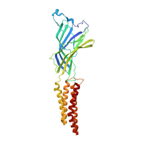
    4IRE

  • PubMed Abstract: 

    The mechanisms of allosteric action within pentameric ligand-gated ion channels (pLGICs) remain to be determined. Using crystallography, site-directed mutagenesis, and two-electrode voltage clamp measurements, we identified two functionally relevant sites in the extracellular (EC) domain of the bacterial pLGIC from Gloeobacter violaceus (GLIC). One site is at the C-loop region, where the NQN mutation (D91N, E177Q, and D178N) eliminated inter-subunit salt bridges in the open-channel GLIC structure and thereby shifted the channel activation to a higher agonist concentration. The other site is below the C-loop, where binding of the anesthetic ketamine inhibited GLIC currents in a concentration dependent manner. To understand how a perturbation signal in the EC domain, either resulting from the NQN mutation or ketamine binding, is transduced to the channel gate, we have used the Perturbation-based Markovian Transmission (PMT) model to determine dynamic responses of the GLIC channel and signaling pathways upon initial perturbations in the EC domain of GLIC. Despite the existence of many possible routes for the initial perturbation signal to reach the channel gate, the PMT model in combination with Yen's algorithm revealed that perturbation signals with the highest probability flow travel either via the β1-β2 loop or through pre-TM1. The β1-β2 loop occurs in either intra- or inter-subunit pathways, while pre-TM1 occurs exclusively in inter-subunit pathways. Residues involved in both types of pathways are well supported by previous experimental data on nAChR. The direct coupling between pre-TM1 and TM2 of the adjacent subunit adds new insight into the allosteric signaling mechanism in pLGICs.


  • Organizational Affiliation
    • Department of Anesthesiology, University of Pittsburgh School of Medicine, Pittsburgh, Pennsylvania, United States of America.

Macromolecule Content 

  • Total Structure Weight: 194.59 kDa 
  • Atom Count: 13,440 
  • Modeled Residue Count: 1,561 
  • Deposited Residue Count: 1,585 
  • Unique protein chains: 1

Macromolecules

Find similar proteins by:|  3D Structure
Entity ID: 1
MoleculeChains  Sequence LengthOrganismDetailsImage
Proton-gated ion channel
A, B, C, D, E
317Gloeobacter violaceus PCC 7421Mutation(s): 3 
Gene Names: glr4197glvI
Membrane Entity: Yes 
UniProt
Find proteins for Q7NDN8 (Gloeobacter violaceus (strain ATCC 29082 / PCC 7421))
Explore Q7NDN8 
Go to UniProtKB:  Q7NDN8
Entity Groups
Sequence Clusters30% Identity50% Identity70% Identity90% Identity95% Identity100% Identity
UniProt GroupQ7NDN8
Sequence Annotations
Expand
Reference Sequence

Small Molecules

Ligands 4 Unique
IDChains Name / Formula / InChI Key2D Diagram3D Interactions
PC1

Query on PC1


Download:Ideal Coordinates CCD File
CA [auth D]
DA [auth D]
G [auth A]
H [auth A]
N [auth B]
CA [auth D],
DA [auth D],
G [auth A],
H [auth A],
N [auth B],
O [auth B],
OA [auth E],
PA [auth E],
V [auth C],
W [auth C]
1,2-DIACYL-SN-GLYCERO-3-PHOSPHOCHOLINE
C44 H88 N O8 P
NRJAVPSFFCBXDT-HUESYALOSA-N
LMD

Query on LMD


Download:Ideal Coordinates CCD File
F [auth A]
KA [auth E]
LA [auth E]
NA [auth E]
T [auth C]
F [auth A],
KA [auth E],
LA [auth E],
NA [auth E],
T [auth C],
U [auth C]
tetradecyl 4-O-alpha-D-glucopyranosyl-beta-D-glucopyranoside
C26 H50 O11
UKPROSIGWJBJGA-IWODYCRQSA-N
OXL

Query on OXL


Download:Ideal Coordinates CCD File
BA [auth D]
EA [auth D]
FA [auth D]
GA [auth D]
HA [auth D]
BA [auth D],
EA [auth D],
FA [auth D],
GA [auth D],
HA [auth D],
I [auth A],
J [auth A],
K [auth A],
MA [auth E],
P [auth B],
Q [auth B],
QA [auth E],
RA [auth E],
SA [auth E],
TA [auth E],
X [auth C],
Y [auth C]
OXALATE ION
C2 O4
MUBZPKHOEPUJKR-UHFFFAOYSA-L
ACT

Query on ACT


Download:Ideal Coordinates CCD File
AA [auth C]
IA [auth D]
JA [auth D]
L [auth A]
M [auth A]
AA [auth C],
IA [auth D],
JA [auth D],
L [auth A],
M [auth A],
R [auth B],
S [auth B],
UA [auth E],
VA [auth E],
Z [auth C]
ACETATE ION
C2 H3 O2
QTBSBXVTEAMEQO-UHFFFAOYSA-M

Experimental Data & Validation

Experimental Data

  • Method: X-RAY DIFFRACTION
  • Resolution: 3.19 Å
  • R-Value Free:  0.243 (Depositor), 0.240 (DCC) 
  • R-Value Work:  0.204 (Depositor), 0.204 (DCC) 
  • R-Value Observed: 0.204 (Depositor) 
Space Group: C 1 2 1
Unit Cell:
Length ( Å )Angle ( ˚ )
a = 182.03α = 90
b = 133.62β = 102.57
c = 161.43γ = 90
Software Package:
Software NamePurpose
XSCALEdata scaling
PHENIXrefinement
PDB_EXTRACTdata extraction
Blu-Icedata collection
XDSdata reduction
XDSdata scaling
PHENIXphasing

Structure Validation

View Full Validation Report


Ligand Structure Quality Assessment 


Entry History 

Deposition Data

Revision History  (Full details and data files)

  • Version 1.0: 2013-04-24
    Type: Initial release
  • Version 1.1: 2013-05-01
    Changes: Structure summary
  • Version 1.2: 2013-05-22
    Changes: Database references
  • Version 1.3: 2023-09-20
    Changes: Data collection, Database references, Derived calculations, Refinement description