4IQL

Crystal Structure of Porphyromonas gingivalis Enoyl-ACP Reductase II (FabK) with cofactors NADPH and FMN


Experimental Data Snapshot

  • Method: X-RAY DIFFRACTION
  • Resolution: 1.94 Å
  • R-Value Free: 0.207 
  • R-Value Work: 0.161 
  • R-Value Observed: 0.163 

Starting Model: experimental
View more details

wwPDB Validation   3D Report Full Report


Ligand Structure Quality Assessment 


This is version 1.4 of the entry. See complete history


Literature

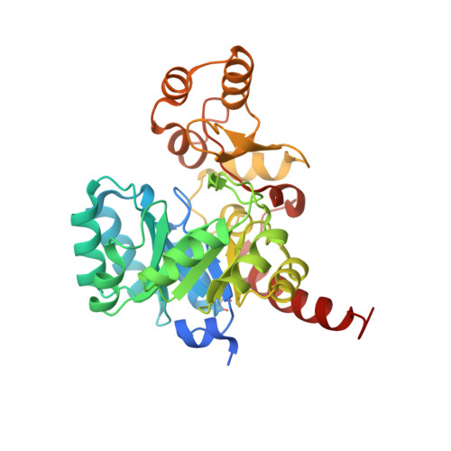
Structural characterization of Porphyromonas gingivalis enoyl-ACP reductase II (FabK).

Hevener, K.E.Santarsiero, B.D.Lee, H.Jones, J.A.Boci, T.Johnson, M.E.Mehboob, S.

(2018) Acta Crystallogr F Struct Biol Commun 74: 105-112

  • DOI: https://doi.org/10.1107/S2053230X18000262
  • Primary Citation of Related Structures:  
    4IQL

  • PubMed Abstract: 

    Enoyl-acyl carrier protein (ACP) reductase II (FabK) is a critical rate-limiting enzyme in the bacterial type II fatty-acid synthesis (FAS II) pathway. FAS II pathway enzymes are markedly disparate from their mammalian analogs in the FAS I pathway in both structure and mechanism. Enzymes involved in bacterial fatty-acid synthesis represent viable drug targets for Gram-negative pathogens, and historical precedent exists for targeting them in the treatment of diseases of the oral cavity. The Gram-negative organism Porphyromonas gingivalis represents a key causative agent of the costly and highly prevalent disease known as chronic periodontitis, and exclusively expresses FabK as its enoyl reductase enzyme in the FAS-II pathway. Together, these characteristics distinguish P. gingivalis FabK (PgFabK) as an attractive and novel narrow-spectrum antibacterial target candidate. PgFabK is a flavoenzyme that is dependent on FMN and NADPH as cofactors for the enzymatic reaction, which reduces the enoyl substrate via a ping-pong mechanism. Here, the structure of the PgFabK enzyme as determined using X-ray crystallography is reported to 1.9 Å resolution with endogenous FMN fully resolved and the NADPH cofactor partially resolved. PgFabK possesses a TIM-barrel motif, and all flexible loops are visible. The determined structure has allowed insight into the structural basis for the NADPH dependence observed in PgFabK and the role of a monovalent cation that has been observed in previous studies to be stringently required for FabK activity. The PgFabK structure and the insights gleaned from its analysis will facilitate structure-based drug-discovery efforts towards the prevention and treatment of P. gingivalis infection.


  • Organizational Affiliation

    Department of Pharmaceutical Sciences, University of Tennessee Health Science Center, 881 Madison Avenue, Suite 571, Memphis, TN 38163-2198, USA.


Macromolecules
Find similar proteins by:  (by identity cutoff)  |  3D Structure
Entity ID: 1
MoleculeChains Sequence LengthOrganismDetailsImage
Enoyl-(Acyl-carrier-protein) reductase II
A, B
333Porphyromonas gingivalis W83Mutation(s): 0 
Gene Names: fabKPG_1416
EC: 1.3.1.9
UniProt
Find proteins for Q7MAW0 (Porphyromonas gingivalis (strain ATCC BAA-308 / W83))
Explore Q7MAW0 
Go to UniProtKB:  Q7MAW0
Entity Groups  
Sequence Clusters30% Identity50% Identity70% Identity90% Identity95% Identity100% Identity
UniProt GroupQ7MAW0
Sequence Annotations
Expand
  • Reference Sequence
Small Molecules
Ligands 4 Unique
IDChains Name / Formula / InChI Key2D Diagram3D Interactions
NDP
Query on NDP

Download Ideal Coordinates CCD File 
D [auth A],
E [auth A],
K [auth B],
L [auth B]
NADPH DIHYDRO-NICOTINAMIDE-ADENINE-DINUCLEOTIDE PHOSPHATE
C21 H30 N7 O17 P3
ACFIXJIJDZMPPO-NNYOXOHSSA-N
FMN
Query on FMN

Download Ideal Coordinates CCD File 
C [auth A],
J [auth B]
FLAVIN MONONUCLEOTIDE
C17 H21 N4 O9 P
FVTCRASFADXXNN-SCRDCRAPSA-N
GOL
Query on GOL

Download Ideal Coordinates CCD File 
F [auth A]GLYCEROL
C3 H8 O3
PEDCQBHIVMGVHV-UHFFFAOYSA-N
NA
Query on NA

Download Ideal Coordinates CCD File 
G [auth A]
H [auth A]
I [auth A]
M [auth B]
N [auth B]
G [auth A],
H [auth A],
I [auth A],
M [auth B],
N [auth B],
O [auth B]
SODIUM ION
Na
FKNQFGJONOIPTF-UHFFFAOYSA-N
Modified Residues  1 Unique
IDChains TypeFormula2D DiagramParent
CSO
Query on CSO
A, B
L-PEPTIDE LINKINGC3 H7 N O3 SCYS
Experimental Data & Validation

Experimental Data

  • Method: X-RAY DIFFRACTION
  • Resolution: 1.94 Å
  • R-Value Free: 0.207 
  • R-Value Work: 0.161 
  • R-Value Observed: 0.163 
  • Space Group: P 21 21 21
Unit Cell:
Length ( Å )Angle ( ˚ )
a = 48.652α = 90
b = 86.652β = 90
c = 150.505γ = 90
Software Package:
Software NamePurpose
HKL-2000data collection
PHASERphasing
REFMACrefinement
XDSdata reduction
XDSdata scaling

Structure Validation

View Full Validation Report


Ligand Structure Quality Assessment 


Entry History 

Deposition Data

Revision History  (Full details and data files)

  • Version 1.0: 2014-01-29
    Type: Initial release
  • Version 1.1: 2018-02-07
    Changes: Database references
  • Version 1.2: 2018-04-18
    Changes: Data collection, Database references
  • Version 1.3: 2023-09-20
    Changes: Data collection, Database references, Derived calculations, Refinement description
  • Version 1.4: 2023-12-06
    Changes: Data collection