4IO2 | pdb_00004io2

Crystal Structure of the AvGluR1 ligand binding domain complex with glutamate at 1.37 Angstrom resolution


Experimental Data Snapshot

  • Method: X-RAY DIFFRACTION
  • Resolution: 1.37 Å
  • R-Value Free: 
    0.169 (Depositor), 0.169 (DCC) 
  • R-Value Work: 
    0.137 (Depositor), 0.137 (DCC) 
  • R-Value Observed: 
    0.138 (Depositor) 

Starting Model: experimental
View more details

wwPDB Validation 3D Report Full Report

Validation slider image for 4IO2

This is version 1.4 of the entry. See complete history

Literature

Anions Mediate Ligand Binding in Adineta vaga Glutamate Receptor Ion Channels.

Lomash, S.Chittori, S.Brown, P.Mayer, M.L.

(2013) Structure 21: 414-425

  • DOI: https://doi.org/10.1016/j.str.2013.01.006
  • Primary Citation Related Structures: 
    4IO2, 4IO3, 4IO4, 4IO5, 4IO6, 4IO7

  • PubMed Abstract: 

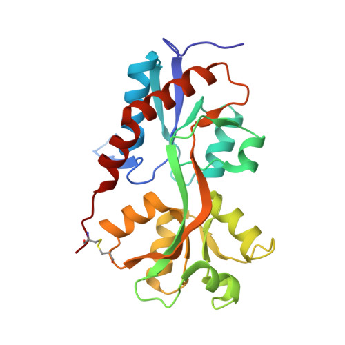
    AvGluR1, a glutamate receptor ion channel from the primitive eukaryote Adineta vaga, is activated by alanine, cysteine, methionine, and phenylalanine, which produce lectin-sensitive desensitizing responses like those to glutamate, aspartate, and serine. AvGluR1 LBD crystal structures reveal an unusual scheme for binding dissimilar ligands that may be utilized by distantly related odorant/chemosensory receptors. Arginine residues in domain 2 coordinate the γ-carboxyl group of glutamate, whereas in the alanine, methionine, and serine complexes a chloride ion acts as a surrogate ligand, replacing the γ-carboxyl group. Removal of Cl(-) lowers affinity for these ligands but not for glutamate or aspartate nor for phenylalanine, which occludes the anion binding site and binds with low affinity. AvGluR1 LBD crystal structures and sedimentation analysis also provide insights into the evolutionary link between prokaryotic and eukaryotic iGluRs and reveal features unique to both classes, emphasizing the need for additional structure-based studies on iGluR-ligand interactions.


  • Organizational Affiliation
    • Laboratory of Cellular and Molecular Neurophysiology, Porter Neuroscience Research Center, National Institute of Child Health and Human Development/NIH/DHHS, Bethesda, MD 20892, USA.

Macromolecule Content 

  • Total Structure Weight: 55.42 kDa 
  • Atom Count: 4,701 
  • Modeled Residue Count: 484 
  • Deposited Residue Count: 496 
  • Unique protein chains: 1

Macromolecules

Find similar proteins by:|  3D Structure
Entity ID: 1
MoleculeChains  Sequence LengthOrganismDetailsImage
Glutamate receptor 1
A, B
248Adineta vagaMutation(s): 0 
Membrane Entity: Yes 
UniProt
Find proteins for E9P5T5 (Adineta vaga)
Explore E9P5T5 
Go to UniProtKB:  E9P5T5
Entity Groups
Sequence Clusters30% Identity50% Identity70% Identity90% Identity95% Identity100% Identity
UniProt GroupE9P5T5
Sequence Annotations
Expand
Reference Sequence

Experimental Data & Validation

Experimental Data

  • Method: X-RAY DIFFRACTION
  • Resolution: 1.37 Å
  • R-Value Free:  0.169 (Depositor), 0.169 (DCC) 
  • R-Value Work:  0.137 (Depositor), 0.137 (DCC) 
  • R-Value Observed: 0.138 (Depositor) 
Space Group: P 1 21 1
Unit Cell:
Length ( Å )Angle ( ˚ )
a = 55.37α = 90
b = 101.052β = 116.46
c = 56.744γ = 90
Software Package:
Software NamePurpose
HKL-2000data collection
PHASERphasing
PHENIXrefinement
HKL-2000data reduction
HKL-2000data scaling

Structure Validation

View Full Validation Report


Entry History 

Deposition Data

Revision History  (Full details and data files)

  • Version 1.0: 2013-02-20
    Type: Initial release
  • Version 1.1: 2013-03-20
    Changes: Database references
  • Version 1.2: 2017-08-09
    Changes: Refinement description, Source and taxonomy
  • Version 1.3: 2023-09-20
    Changes: Data collection, Database references, Derived calculations, Refinement description
  • Version 1.4: 2024-11-27
    Changes: Structure summary