4IL3 | pdb_00004il3

Crystal Structure of S. mikatae Ste24p


Experimental Data Snapshot

  • Method: X-RAY DIFFRACTION
  • Resolution: 3.10 Å
  • R-Value Free: 
    0.293 (Depositor), 0.281 (DCC) 
  • R-Value Work: 
    0.270 (Depositor), 0.282 (DCC) 
  • R-Value Observed: 
    0.271 (Depositor) 

Starting Model: experimental
View more details

wwPDB Validation 3D Report Full Report

Validation slider image for 4IL3

This is version 1.4 of the entry. See complete history

Literature

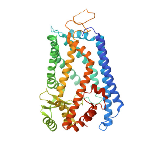
Structure of the integral membrane protein CAAX protease Ste24p.

Pryor, E.E.Horanyi, P.S.Clark, K.M.Fedoriw, N.Connelly, S.M.Koszelak-Rosenblum, M.Zhu, G.Malkowski, M.G.Wiener, M.C.Dumont, M.E.

(2013) Science 339: 1600-1604

  • DOI: https://doi.org/10.1126/science.1232048
  • Primary Citation Related Structures: 
    4IL3

  • PubMed Abstract: 

    Posttranslational lipidation provides critical modulation of the functions of some proteins. Isoprenoids (i.e., farnesyl or geranylgeranyl groups) are attached to cysteine residues in proteins containing C-terminal CAAX sequence motifs (where A is an aliphatic residue and X is any residue). Isoprenylation is followed by cleavage of the AAX amino acid residues and, in some cases, by additional proteolytic cuts. We determined the crystal structure of the CAAX protease Ste24p, a zinc metalloprotease catalyzing two proteolytic steps in the maturation of yeast mating pheromone a-factor. The Ste24p core structure is a ring of seven transmembrane helices enclosing a voluminous cavity containing the active site and substrate-binding groove. The cavity is accessible to the external milieu by means of gaps between splayed transmembrane helices. We hypothesize that cleavage proceeds by means of a processive mechanism of substrate insertion, translocation, and ejection.


  • Organizational Affiliation
    • Membrane Protein Structural Biology Consortium, USA.

Macromolecule Content 

  • Total Structure Weight: 106.81 kDa 
  • Atom Count: 6,975 
  • Modeled Residue Count: 853 
  • Deposited Residue Count: 922 
  • Unique protein chains: 1

Macromolecules

Find similar proteins by:|  3D Structure
Entity ID: 1
MoleculeChains  Sequence LengthOrganismDetailsImage
Ste24p
A, B
461Saccharomyces mikataeMutation(s): 0 
EC: 3.4.24.84
Membrane Entity: Yes 
UniProt
Find proteins for M4GGS2 (Saccharomyces mikatae)
Explore M4GGS2 
Go to UniProtKB:  M4GGS2
Entity Groups
Sequence Clusters30% Identity50% Identity70% Identity90% Identity95% Identity100% Identity
UniProt GroupM4GGS2
Sequence Annotations
Expand
Reference Sequence

Experimental Data & Validation

Experimental Data

  • Method: X-RAY DIFFRACTION
  • Resolution: 3.10 Å
  • R-Value Free:  0.293 (Depositor), 0.281 (DCC) 
  • R-Value Work:  0.270 (Depositor), 0.282 (DCC) 
  • R-Value Observed: 0.271 (Depositor) 
Space Group: P 65
Unit Cell:
Length ( Å )Angle ( ˚ )
a = 143.53α = 90
b = 143.53β = 90
c = 197.024γ = 120
Software Package:
Software NamePurpose
DENZOdata reduction
SCALEPACKdata scaling
PHASERphasing
RESOLVEphasing
PHENIXrefinement
PDB_EXTRACTdata extraction
MAR345data collection
PHENIXphasing

Structure Validation

View Full Validation Report


Entry History 

Revision History  (Full details and data files)

  • Version 1.0: 2013-02-20
    Type: Initial release
  • Version 1.1: 2013-04-03
    Changes: Database references
  • Version 1.2: 2013-06-19
    Changes: Database references
  • Version 1.3: 2017-11-15
    Changes: Refinement description
  • Version 1.4: 2023-09-20
    Changes: Data collection, Database references, Derived calculations, Refinement description