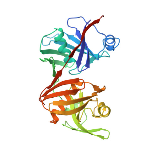
4IK0

Crystal structure of diaminopimelate epimerase Y268A mutant from Escherichia coli


Experimental Data Snapshot

  • Method: X-RAY DIFFRACTION
  • Resolution: 2.05 Å
  • R-Value Free: 0.217 
  • R-Value Work: 0.176 
  • R-Value Observed: 0.178 

wwPDB Validation   3D Report Full Report


This is version 1.3 of the entry. See complete history


Literature

Dimerization of bacterial diaminopimelate epimerase is essential for catalysis

Hor, L.Dobson, R.C.J.Downton, M.T.Wagner, J.Hutton, C.A.Perugini, M.A.

(2013) J Biol Chem 288: 9238-9248

  • DOI: https://doi.org/10.1074/jbc.M113.450148
  • Primary Citation of Related Structures:  
    4IJZ, 4IK0

  • PubMed Abstract: 

    Diaminopimelate (DAP) epimerase is involved in the biosynthesis of meso-DAP and lysine, which are important precursors for the synthesis of peptidoglycan, housekeeping proteins, and virulence factors in bacteria. Accordingly, DAP epimerase is a promising antimicrobial target. Previous studies report that DAP epimerase exists as a monomeric enzyme. However, we show using analytical ultracentrifugation, X-ray crystallography, and enzyme kinetic analyses that DAP epimerase from Escherichia coli exists as a functional dimer in solution and the crystal state. Furthermore, the 2.0-Å X-ray crystal structure of the E. coli DAP epimerase dimer shows for the first time that the enzyme exists in an open, active conformation. The importance of dimerization was subsequently probed by using site-directed mutagenesis to generate a monomeric mutant (Y268A). Our studies show that Y268A is catalytically inactive, thus demonstrating that dimerization of DAP epimerase is essential for catalysis. Molecular dynamics simulations indicate that the DAP epimerase monomer is inherently more flexible than the dimer, suggesting that dimerization optimizes protein dynamics to support function. Our findings offer insight into the development of novel antimicrobial agents targeting the dimeric antibiotic target DAP epimerase.


  • Organizational Affiliation

    Department of Biochemistry, La Trobe Institute for Molecular Science, La Trobe University, Melbourne, Victoria 3086, Australia.


Macromolecules
Find similar proteins by:  (by identity cutoff)  |  3D Structure
Entity ID: 1
MoleculeChains Sequence LengthOrganismDetailsImage
Diaminopimelate epimerase
A, B
280Escherichia coli K-12Mutation(s): 1 
Gene Names: dapF
EC: 5.1.1.7
UniProt
Find proteins for P0A6K1 (Escherichia coli (strain K12))
Explore P0A6K1 
Go to UniProtKB:  P0A6K1
Entity Groups  
Sequence Clusters30% Identity50% Identity70% Identity90% Identity95% Identity100% Identity
UniProt GroupP0A6K1
Sequence Annotations
Expand
  • Reference Sequence
Small Molecules
Ligands 1 Unique
IDChains Name / Formula / InChI Key2D Diagram3D Interactions
IOD
Query on IOD

Download Ideal Coordinates CCD File 
C [auth A]
D [auth A]
E [auth A]
F [auth A]
G [auth A]
C [auth A],
D [auth A],
E [auth A],
F [auth A],
G [auth A],
H [auth A],
I [auth B],
J [auth B],
K [auth B],
L [auth B],
M [auth B],
N [auth B],
O [auth B]
IODIDE ION
I
XMBWDFGMSWQBCA-UHFFFAOYSA-M
Experimental Data & Validation

Experimental Data

  • Method: X-RAY DIFFRACTION
  • Resolution: 2.05 Å
  • R-Value Free: 0.217 
  • R-Value Work: 0.176 
  • R-Value Observed: 0.178 
  • Space Group: P 41 21 2
Unit Cell:
Length ( Å )Angle ( ˚ )
a = 89.18α = 90
b = 89.18β = 90
c = 179.45γ = 90
Software Package:
Software NamePurpose
SCALAdata scaling
PHASERphasing
REFMACrefinement
PDB_EXTRACTdata extraction
MOSFLMdata reduction

Structure Validation

View Full Validation Report


Entry History 

Deposition Data

Revision History  (Full details and data files)

  • Version 1.0: 2013-02-20
    Type: Initial release
  • Version 1.1: 2013-03-06
    Changes: Database references
  • Version 1.2: 2013-04-17
    Changes: Database references
  • Version 1.3: 2024-03-20
    Changes: Data collection, Database references, Derived calculations