4IH3 | pdb_00004ih3

2.5 Angstroms X-ray crystal structure of of human 2-amino-3-carboxymuconate-6-semialdehyde decarboxylase in complex with dipicolinic acid


Experimental Data Snapshot

  • Method: X-RAY DIFFRACTION
  • Resolution: 2.49 Å
  • R-Value Free: 
    0.283 (Depositor), 0.279 (DCC) 
  • R-Value Work: 
    0.198 (Depositor), 0.192 (DCC) 
  • R-Value Observed: 
    0.202 (Depositor) 

Starting Model: experimental
View more details

wwPDB Validation 3D Report Full Report

Validation slider image for 4IH3

Ligand Structure Quality Assessment 


This is version 1.3 of the entry. See complete history

Literature

Human alpha-amino-beta-carboxymuconate-epsilon-semialdehyde decarboxylase (ACMSD): A structural and mechanistic unveiling.

Huo, L.Liu, F.Iwaki, H.Li, T.Hasegawa, Y.Liu, A.

(2015) Proteins 83: 178-187

  • DOI: https://doi.org/10.1002/prot.24722
  • Primary Citation Related Structures: 
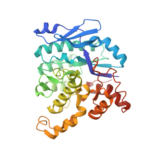
    4IGN, 4IH3, 4OFC

  • PubMed Abstract: 

    Human α-amino-β-carboxymuconate-ε-semialdehyde decarboxylase determines the fate of tryptophan metabolites in the kynurenine pathway by controlling the quinolinate levels for de novo nicotinamide adenine dinucleotide biosynthesis. The unstable nature of its substrate has made gaining insight into its reaction mechanism difficult. Our electron paramagnetic resonance (EPR) spectroscopic study on the Cu-substituted human enzyme suggests that the native substrate does not directly ligate to the metal ion. Substrate binding did not result in a change of either the hyperfine structure or the super-hyperfine structure of the EPR spectrum. We also determined the crystal structure of the human enzyme in its native catalytically active state (at 1.99 Å resolution), a substrate analogue-bound form (2.50 Å resolution), and a selected active site mutant form with one of the putative substrate binding residues altered (2.32 Å resolution). These structures illustrate that each asymmetric unit contains three pairs of dimers. Consistent with the EPR findings, the ligand-bound complex structure shows that the substrate analogue does not directly coordinate to the metal ion but is bound to the active site by two arginine residues through noncovalent interactions.


  • Organizational Affiliation
    • Department of Chemistry and the Center for Diagnostics and Therapeutics, Georgia State University, Atlanta, Georgia 30303.

Macromolecule Content 

  • Total Structure Weight: 226.7 kDa 
  • Atom Count: 16,174 
  • Modeled Residue Count: 1,992 
  • Deposited Residue Count: 1,992 
  • Unique protein chains: 1

Macromolecules

Find similar proteins by:|  3D Structure
Entity ID: 1
MoleculeChains  Sequence LengthOrganismDetailsImage
2-amino-3-carboxymuconate-6-semialdehyde decarboxylase
A, B, C, D, E
A, B, C, D, E, F
332Homo sapiensMutation(s): 0 
Gene Names: ACMSD
EC: 4.1.1.45
UniProt & NIH Common Fund Data Resources
Find proteins for Q8TDX5 (Homo sapiens)
Explore Q8TDX5 
Go to UniProtKB:  Q8TDX5
PHAROS:  Q8TDX5
GTEx:  ENSG00000153086 
Entity Groups
Sequence Clusters30% Identity50% Identity70% Identity90% Identity95% Identity100% Identity
UniProt GroupQ8TDX5
Sequence Annotations
Expand
Reference Sequence

Small Molecules

Ligands 2 Unique
IDChains Name / Formula / InChI Key2D Diagram3D Interactions
PDC

Query on PDC


Download:Ideal Coordinates CCD File
H [auth A]
J [auth B]
L [auth C]
N [auth D]
P [auth E]
H [auth A],
J [auth B],
L [auth C],
N [auth D],
P [auth E],
R [auth F]
PYRIDINE-2,6-DICARBOXYLIC ACID
C7 H5 N O4
WJJMNDUMQPNECX-UHFFFAOYSA-N
ZN

Query on ZN


Download:Ideal Coordinates CCD File
G [auth A]
I [auth B]
K [auth C]
M [auth D]
O [auth E]
G [auth A],
I [auth B],
K [auth C],
M [auth D],
O [auth E],
Q [auth F]
ZINC ION
Zn
PTFCDOFLOPIGGS-UHFFFAOYSA-N
Binding Affinity Annotations 
IDSourceBinding Affinity
PDC BindingDB:  4IH3 Ki: 1.52e+4 (nM) from 1 assay(s)
IC50: 1.52e+4 (nM) from 1 assay(s)

Experimental Data & Validation

Experimental Data

  • Method: X-RAY DIFFRACTION
  • Resolution: 2.49 Å
  • R-Value Free:  0.283 (Depositor), 0.279 (DCC) 
  • R-Value Work:  0.198 (Depositor), 0.192 (DCC) 
  • R-Value Observed: 0.202 (Depositor) 
Space Group: P 21 21 21
Unit Cell:
Length ( Å )Angle ( ˚ )
a = 88.368α = 90
b = 100.655β = 90
c = 233.117γ = 90
Software Package:
Software NamePurpose
SCALEPACKdata scaling
PHENIXrefinement
PDB_EXTRACTdata extraction
MAR345dtbdata collection
HKL-2000data reduction
MOLREPphasing

Structure Validation

View Full Validation Report


Ligand Structure Quality Assessment 


Entry History 

Deposition Data

Revision History  (Full details and data files)

  • Version 1.0: 2014-05-21
    Type: Initial release
  • Version 1.1: 2014-11-26
    Changes: Database references
  • Version 1.2: 2014-12-31
    Changes: Database references
  • Version 1.3: 2023-09-20
    Changes: Data collection, Database references, Derived calculations, Refinement description