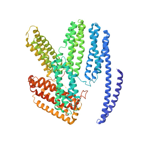
4IGG

Full-length human alpha-catenin crystal structure


Experimental Data Snapshot

  • Method: X-RAY DIFFRACTION
  • Resolution: 3.66 Å
  • R-Value Free: 0.241 
  • R-Value Work: 0.217 
  • R-Value Observed: 0.219 

wwPDB Validation   3D Report Full Report


This is version 1.2 of the entry. See complete history


Literature

Dimer asymmetry defines alpha-catenin interactions.

Rangarajan, E.S.Izard, T.

(2013) Nat Struct Mol Biol 20: 188-193

  • DOI: https://doi.org/10.1038/nsmb.2479
  • Primary Citation of Related Structures:  
    4IGG

  • PubMed Abstract: 

    The F-actin-binding cytoskeletal protein α-catenin interacts with β-catenin-cadherin complexes and stabilizes cell-cell junctions. The β-catenin-α-catenin complex cannot bind F-actin, whereas interactions of α-catenin with the cytoskeletal protein vinculin appear to be necessary to stabilize adherens junctions. Here we report the crystal structure of nearly full-length human α-catenin at 3.7-Å resolution. α-catenin forms an asymmetric dimer where the four-helix bundle domains of each subunit engage in distinct intermolecular interactions. This results in a left handshake-like dimer, wherein the two subunits have remarkably different conformations. The crystal structure explains why dimeric α-catenin has a higher affinity for F-actin than does monomeric α-catenin, why the β-catenin-α-catenin complex does not bind F-actin, how activated vinculin links the cadherin-catenin complex to the cytoskeleton and why α-catenin but not inactive vinculin can bind F-actin.


  • Organizational Affiliation

    Department of Cancer Biology, Scripps Research Institute, Jupiter, Florida, USA.


Macromolecules
Find similar proteins by:  (by identity cutoff)  |  3D Structure
Entity ID: 1
MoleculeChains Sequence LengthOrganismDetailsImage
Catenin alpha-1
A, B
832Homo sapiensMutation(s): 0 
Gene Names: CTNNA1
UniProt & NIH Common Fund Data Resources
Find proteins for P35221 (Homo sapiens)
Explore P35221 
Go to UniProtKB:  P35221
PHAROS:  P35221
GTEx:  ENSG00000044115 
Entity Groups  
Sequence Clusters30% Identity50% Identity70% Identity90% Identity95% Identity100% Identity
UniProt GroupP35221
Sequence Annotations
Expand
  • Reference Sequence
Experimental Data & Validation

Experimental Data

  • Method: X-RAY DIFFRACTION
  • Resolution: 3.66 Å
  • R-Value Free: 0.241 
  • R-Value Work: 0.217 
  • R-Value Observed: 0.219 
  • Space Group: P 32
Unit Cell:
Length ( Å )Angle ( ˚ )
a = 145.629α = 90
b = 145.629β = 90
c = 139.081γ = 120
Software Package:
Software NamePurpose
SCALAdata scaling
SHARPphasing
SOLOMONphasing
TNTrefinement
PDB_EXTRACTdata extraction
SERGUIdata collection
XSCALEdata scaling
BUSTERrefinement

Structure Validation

View Full Validation Report


Entry History 

Deposition Data

Revision History  (Full details and data files)

  • Version 1.0: 2012-12-26
    Type: Initial release
  • Version 1.1: 2013-09-11
    Changes: Database references
  • Version 1.2: 2024-02-28
    Changes: Data collection, Database references, Derived calculations