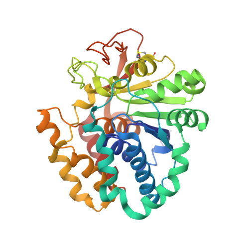
4I73

Crystal structure of the Trypanosoma brucei Inosine-Adenosine-Guanosine nucleoside hydrolase in complex with compound UAMC-00312


Experimental Data Snapshot

  • Method: X-RAY DIFFRACTION
  • Resolution: 2.18 Å
  • R-Value Free: 0.248 
  • R-Value Work: 0.200 
  • R-Value Observed: 0.203 

wwPDB Validation   3D Report Full Report


Ligand Structure Quality Assessment 


This is version 1.2 of the entry. See complete history


Literature

Structures of purine nucleosidase from Trypanosoma brucei bound to isozyme-specific trypanocidals and a novel metalorganic inhibitor

Giannese, F.Berg, M.Van der Veken, P.Castagna, V.Tornaghi, P.Augustyns, K.Degano, M.

(2013) Acta Crystallogr D Biol Crystallogr 69: 1553-1566

  • DOI: https://doi.org/10.1107/S0907444913010792
  • Primary Citation of Related Structures:  
    4I70, 4I71, 4I72, 4I73, 4I74, 4I75

  • PubMed Abstract: 

    Sleeping sickness is a deadly disease that primarily affects sub-Saharan Africa and is caused by protozoan parasites of the Trypanosoma genus. Trypanosomes are purine auxotrophs and their uptake pathway has long been appreciated as an attractive target for drug design. Recently, one tight-binding competitive inhibitor of the trypanosomal purine-specific nucleoside hydrolase (IAGNH) showed remarkable trypanocidal activity in a murine model of infection. Here, the enzymatic characterization of T. brucei brucei IAGNH is presented, together with its high-resolution structures in the unliganded form and in complexes with different inhibitors, including the trypanocidal compound UAMC-00363. A description of the crucial contacts that account for the high-affinity inhibition of IAGNH by iminoribitol-based compounds is provided and the molecular mechanism underlying the conformational change necessary for enzymatic catalysis is identified. It is demonstrated for the first time that metalorganic complexes can compete for binding at the active site of nucleoside hydrolase enzymes, mimicking the positively charged transition state of the enzymatic reaction. Moreover, we show that divalent metal ions can act as noncompetitive IAGNH inhibitors, stabilizing a nonproductive conformation of the catalytic loop. These results open a path for rational improvement of the potency and the selectivity of existing compounds and suggest new scaffolds that may be used as blueprints for the design of novel antitrypanosomal compounds.


  • Organizational Affiliation

    Biocrystallography Unit, Department of Immunology, Transplantation and Infectious Diseases, Scientific Institute San Raffaele, via Olgettina 58, 20132 Milano, Italy.


Macromolecules
Find similar proteins by:  (by identity cutoff)  |  3D Structure
Entity ID: 1
MoleculeChains Sequence LengthOrganismDetailsImage
Inosine-adenosine-guanosine-nucleoside hydrolase
A, B, C, D
330Trypanosoma brucei brucei TREU927Mutation(s): 0 
Gene Names: Tb927.3.2960
EC: 3.2.2.1
UniProt
Find proteins for Q57ZL6 (Trypanosoma brucei brucei (strain 927/4 GUTat10.1))
Explore Q57ZL6 
Go to UniProtKB:  Q57ZL6
Entity Groups  
Sequence Clusters30% Identity50% Identity70% Identity90% Identity95% Identity100% Identity
UniProt GroupQ57ZL6
Sequence Annotations
Expand
  • Reference Sequence
Small Molecules
Ligands 3 Unique
IDChains Name / Formula / InChI Key2D Diagram3D Interactions
MBY
Query on MBY

Download Ideal Coordinates CCD File 
F [auth A],
N [auth B],
R [auth C],
Z [auth D]
(2R,3R,4S)-2-(hydroxymethyl)-1-[(4-hydroxythieno[3,2-d]pyrimidin-7-yl)methyl]pyrrolidine-3,4-diol
C12 H15 N3 O4 S
UZJHCHSHAYEOTK-KHQFGBGNSA-N
NI
Query on NI

Download Ideal Coordinates CCD File 
AA [auth D]
BA [auth D]
CA [auth D]
G [auth A]
H [auth A]
AA [auth D],
BA [auth D],
CA [auth D],
G [auth A],
H [auth A],
I [auth A],
J [auth A],
K [auth A],
L [auth A],
O [auth B],
P [auth B],
S [auth C],
T [auth C],
U [auth C],
V [auth C],
W [auth C],
X [auth C]
NICKEL (II) ION
Ni
VEQPNABPJHWNSG-UHFFFAOYSA-N
CA
Query on CA

Download Ideal Coordinates CCD File 
E [auth A],
M [auth B],
Q [auth C],
Y [auth D]
CALCIUM ION
Ca
BHPQYMZQTOCNFJ-UHFFFAOYSA-N
Binding Affinity Annotations 
IDSourceBinding Affinity
MBY PDBBind:  4I73 Ki: 190 (nM) from 1 assay(s)
Experimental Data & Validation

Experimental Data

  • Method: X-RAY DIFFRACTION
  • Resolution: 2.18 Å
  • R-Value Free: 0.248 
  • R-Value Work: 0.200 
  • R-Value Observed: 0.203 
  • Space Group: P 1 21 1
Unit Cell:
Length ( Å )Angle ( ˚ )
a = 63.17α = 90
b = 131.67β = 91.33
c = 71.85γ = 90
Software Package:
Software NamePurpose
XSCALEdata scaling
REFMACrefinement
PDB_EXTRACTdata extraction
DNAdata collection
XDSdata reduction
MOLREPphasing

Structure Validation

View Full Validation Report


Ligand Structure Quality Assessment 


Entry History 

Deposition Data

Revision History  (Full details and data files)

  • Version 1.0: 2013-08-07
    Type: Initial release
  • Version 1.1: 2013-12-18
    Changes: Database references
  • Version 1.2: 2024-10-30
    Changes: Data collection, Database references, Derived calculations, Refinement description, Structure summary