4I52

scMenB im complex with 1-hydroxy-2-naphthoyl-CoA


Experimental Data Snapshot

  • Method: X-RAY DIFFRACTION
  • Resolution: 2.35 Å
  • R-Value Free: 0.256 
  • R-Value Work: 0.229 
  • R-Value Observed: 0.230 

wwPDB Validation   3D Report Full Report


Ligand Structure Quality Assessment 


This is version 1.2 of the entry. See complete history


Literature

Structural basis of the induced-fit mechanism of 1,4-dihydroxy-2-naphthoyl coenzyme a synthase from the crotonase fold superfamily

Sun, Y.Song, H.Li, J.Li, Y.Jiang, M.Zhou, J.Guo, Z.

(2013) PLoS One 8: e63095-e63095

  • DOI: https://doi.org/10.1371/journal.pone.0063095
  • Primary Citation of Related Structures:  
    4I42, 4I4Z, 4I52

  • PubMed Abstract: 

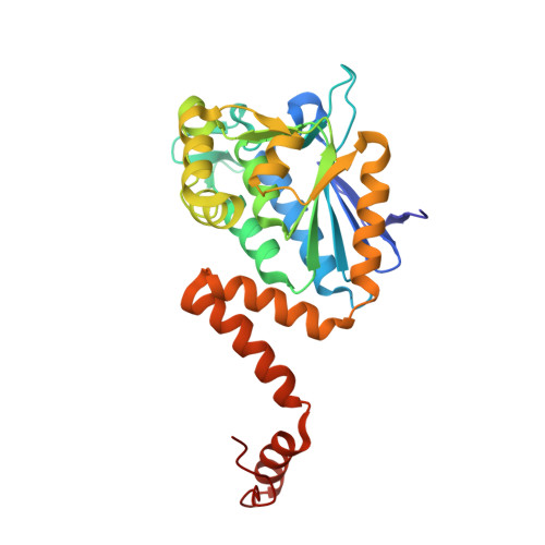
    1, 4-Dihydroxy-2-naphthoyl coenzyme A (DHNA-CoA) synthase is a typical crotonase fold enzyme with an implicated role of conformational changes in catalysis. We have identified these conformational changes by determining the structures of its Escherichia coli and Synechocystis sp. PCC6803 orthologues in complex with a product analog. The structural changes include the folding of an active-site loop into a β-hairpin and significant reorientation of a helix at the carboxy terminus. Interestingly, a new interface is formed between the ordered loop and the reoriented helix, both of which also form additional interactions with the coenzyme A moiety of the ligand. Site-directed mutation of the amino acid residues involved in these ligand-induced interactions significantly diminishes the enzyme activity. These results suggest a catalytically essential induced-fit that is likely initiated by the enzyme-ligand interactions at the active site.


  • Organizational Affiliation

    Department of Chemistry and State Key Laboratory of Molecular Neuroscience, The Hong Kong University of Science and Technology, Clear Water Bay, Kowloon, Hong Kong SAR, China.


Macromolecules
Find similar proteins by:  (by identity cutoff)  |  3D Structure
Entity ID: 1
MoleculeChains Sequence LengthOrganismDetailsImage
Naphthoate synthase
A, B, C, D, E
A, B, C, D, E, F, G, H, I
275Synechocystis sp. PCC 6803 substr. KazusaMutation(s): 0 
EC: 4.1.3.36
UniProt
Find proteins for P73495 (Synechocystis sp. (strain PCC 6803 / Kazusa))
Explore P73495 
Go to UniProtKB:  P73495
Entity Groups  
Sequence Clusters30% Identity50% Identity70% Identity90% Identity95% Identity100% Identity
UniProt GroupP73495
Sequence Annotations
Expand
  • Reference Sequence
Small Molecules
Ligands 2 Unique
IDChains Name / Formula / InChI Key2D Diagram3D Interactions
1HA
Query on 1HA

Download Ideal Coordinates CCD File 
J [auth A]
L [auth B]
N [auth C]
P [auth D]
R [auth E]
J [auth A],
L [auth B],
N [auth C],
P [auth D],
R [auth E],
T [auth F],
V [auth G],
X [auth H],
Z [auth I]
1-hydroxy-2-naphthoyl-CoA
C32 H42 N7 O18 P3 S
ZHSLHXZSNOSMQG-HSJNEKGZSA-N
CL
Query on CL

Download Ideal Coordinates CCD File 
AA [auth I]
K [auth A]
M [auth B]
O [auth C]
Q [auth D]
AA [auth I],
K [auth A],
M [auth B],
O [auth C],
Q [auth D],
S [auth E],
U [auth F],
W [auth G],
Y [auth H]
CHLORIDE ION
Cl
VEXZGXHMUGYJMC-UHFFFAOYSA-M
Experimental Data & Validation

Experimental Data

  • Method: X-RAY DIFFRACTION
  • Resolution: 2.35 Å
  • R-Value Free: 0.256 
  • R-Value Work: 0.229 
  • R-Value Observed: 0.230 
  • Space Group: P 32 2 1
Unit Cell:
Length ( Å )Angle ( ˚ )
a = 138.843α = 90
b = 138.843β = 90
c = 221.217γ = 120
Software Package:
Software NamePurpose
HKL-2000data collection
PHASERphasing
REFMACrefinement
HKL-2000data reduction
HKL-2000data scaling

Structure Validation

View Full Validation Report


Ligand Structure Quality Assessment 


Entry History 

Deposition Data

Revision History  (Full details and data files)

  • Version 1.0: 2013-05-08
    Type: Initial release
  • Version 1.1: 2013-06-19
    Changes: Database references
  • Version 1.2: 2023-11-08
    Changes: Data collection, Database references, Derived calculations, Refinement description