4I0O | pdb_00004i0o

Nucleoporin ELYS (aa1-494), Mus musculus


Experimental Data Snapshot

  • Method: X-RAY DIFFRACTION
  • Resolution: 1.90 Å
  • R-Value Free: 
    0.214 (Depositor), 0.215 (DCC) 
  • R-Value Work: 
    0.180 (Depositor), 0.182 (DCC) 
  • R-Value Observed: 
    0.182 (Depositor) 

wwPDB Validation 3D Report Full Report

Validation slider image for 4I0O

This is version 1.1 of the entry. See complete history

Literature

Structural and Functional Studies of the 252 kDa Nucleoporin ELYS Reveal Distinct Roles for Its Three Tethered Domains.

Bilokapic, S.Schwartz, T.U.

(2013) Structure 21: 572-580

  • DOI: https://doi.org/10.1016/j.str.2013.02.006
  • Primary Citation Related Structures: 
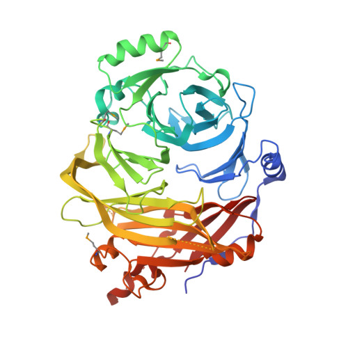
    4I0O

  • PubMed Abstract: 

    In metazoa, the nuclear envelope (NE), together with the embedded nuclear pore complexes (NPCs), breaks down and reassembles during cell division. It is suggested that ELYS, a nucleoporin, binds to chromatin in an initial step of postmitotic NPC assembly and subsequently recruits the essential Y-subcomplex, the major scaffolding unit of the NPC. Here, we show that ELYS contains three domains: an N-terminal β-propeller domain, a central α-helical domain, and a C-terminal disordered region. While the disordered region is responsible for the interactions with chromatin, the two preceding domains synergistically mediate tethering to the NPC. We present the crystal structure of the seven-bladed β-propeller domain at 1.9 Å resolution. Analysis of the β-propeller surface reveals the regions that are required for NPC anchorage. We discuss the possible roles of ELYS in the context of the NPC scaffold architecture.


  • Organizational Affiliation
    • Department of Biology, Massachusetts Institute of Technology, 77 Massachusetts Avenue, Cambridge, MA 02139, USA.

Macromolecule Content 

  • Total Structure Weight: 55.03 kDa 
  • Atom Count: 3,798 
  • Modeled Residue Count: 463 
  • Deposited Residue Count: 497 
  • Unique protein chains: 1

Macromolecules

Find similar proteins by:|  3D Structure
Entity ID: 1
MoleculeChains  Sequence LengthOrganismDetailsImage
Protein ELYS497Mus musculusMutation(s): 0 
Gene Names: Ahctf1Elys
UniProt & NIH Common Fund Data Resources
Find proteins for Q8CJF7 (Mus musculus)
Explore Q8CJF7 
Go to UniProtKB:  Q8CJF7
IMPC:  MGI:1915033
Entity Groups
Sequence Clusters30% Identity50% Identity70% Identity90% Identity95% Identity100% Identity
UniProt GroupQ8CJF7
Sequence Annotations
Expand
Reference Sequence

Small Molecules

Modified Residues  1 Unique
IDChains TypeFormula2D DiagramParent
MSE
Query on MSE
A
L-PEPTIDE LINKINGC5 H11 N O2 SeMET

Experimental Data & Validation

Experimental Data

  • Method: X-RAY DIFFRACTION
  • Resolution: 1.90 Å
  • R-Value Free:  0.214 (Depositor), 0.215 (DCC) 
  • R-Value Work:  0.180 (Depositor), 0.182 (DCC) 
  • R-Value Observed: 0.182 (Depositor) 
Space Group: P 1 21 1
Unit Cell:
Length ( Å )Angle ( ˚ )
a = 52.598α = 90
b = 78.117β = 106.59
c = 58.849γ = 90
Software Package:
Software NamePurpose
SCALEPACKdata scaling
PHENIXrefinement
PDB_EXTRACTdata extraction
HKL-2000data collection
HKL-2000data reduction
SHARPphasing

Structure Validation

View Full Validation Report


Entry History 

Deposition Data

Revision History  (Full details and data files)

  • Version 1.0: 2013-04-17
    Type: Initial release
  • Version 1.1: 2024-11-20
    Changes: Data collection, Database references, Derived calculations, Structure summary