4HYW | pdb_00004hyw

Pyruvate kinase (PYK) from Trypanosoma brucei in the presence of Magnesium and F26BP


Experimental Data Snapshot

  • Method: X-RAY DIFFRACTION
  • Resolution: 2.35 Å
  • R-Value Free: 
    0.199 (Depositor), 0.199 (DCC) 
  • R-Value Work: 
    0.164 (Depositor), 0.163 (DCC) 
  • R-Value Observed: 
    0.166 (Depositor) 

wwPDB Validation 3D Report Full Report

Validation slider image for 4HYW

Ligand Structure Quality Assessment 


This is version 1.2 of the entry. See complete history

Literature

`In crystallo' substrate binding triggers major domain movements and reveals magnesium as a co-activator of Trypanosoma brucei pyruvate kinase.

Zhong, W.Morgan, H.P.McNae, I.W.Michels, P.A.Fothergill-Gilmore, L.A.Walkinshaw, M.D.

(2013) Acta Crystallogr D Biol Crystallogr 69: 1768-1779

  • DOI: https://doi.org/10.1107/S0907444913013875
  • Primary Citation Related Structures: 
    4HYV, 4HYW

  • PubMed Abstract: 

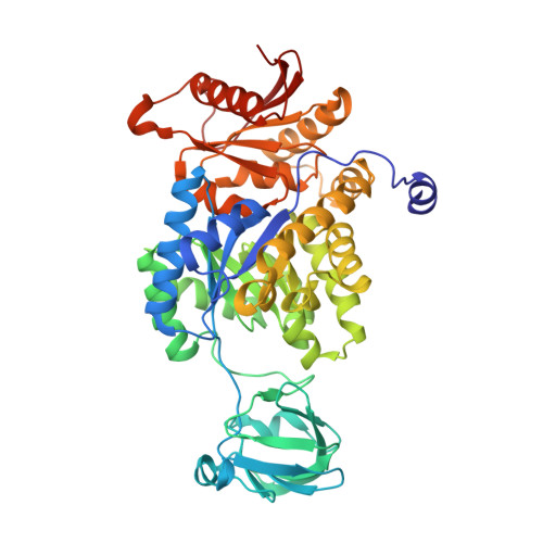
    The active site of pyruvate kinase (PYK) is located between the AC core of the enzyme and a mobile lid corresponding to domain B. Many PYK structures have already been determined, but the first `effector-only' structure and the first with PEP (the true natural substrate) are now reported for the enzyme from Trypanosoma brucei. PEP soaked into crystals of the enzyme with bound allosteric activator fructose 2,6-bisphosphate (F26BP) and Mg(2+) triggers a substantial 23° rotation of the B domain `in crystallo', resulting in a partially closed active site. The interplay of side chains with Mg(2+) and PEP may explain the mechanism of the domain movement. Furthermore, it is apparent that when F26BP is present but PEP is absent Mg(2+) occupies a position that is distinct from the two canonical Mg(2+)-binding sites at the active site. This third site is adjacent to the active site and involves the same amino-acid side chains as in canonical site 1 but in altered orientations. Site 3 acts to sequester Mg(2+) in a `priming' position such that the enzyme is maintained in its R-state conformation. In this way, Mg(2+) cooperates with F26BP to ensure that the enzyme is in a conformation that has a high affinity for the substrate.


  • Organizational Affiliation
    • Centre for Translational and Chemical Biology, School of Biological Sciences, The University of Edinburgh, Mayfield Road, Edinburgh EH9 3JR, Scotland.

Macromolecule Content 

  • Total Structure Weight: 110.01 kDa 
  • Atom Count: 8,289 
  • Modeled Residue Count: 994 
  • Deposited Residue Count: 998 
  • Unique protein chains: 1

Macromolecules

Find similar proteins by:|  3D Structure
Entity ID: 1
MoleculeChains  Sequence LengthOrganismDetailsImage
Pyruvate kinase 1A [auth B],
B [auth A]
499Trypanosoma brucei bruceiMutation(s): 0 
Gene Names: PYK1
EC: 2.7.1.40
UniProt
Find proteins for P30615 (Trypanosoma brucei brucei)
Explore P30615 
Go to UniProtKB:  P30615
Entity Groups
Sequence Clusters30% Identity50% Identity70% Identity90% Identity95% Identity100% Identity
UniProt GroupP30615
Sequence Annotations
Expand
Reference Sequence

Small Molecules

Ligands 5 Unique
IDChains Name / Formula / InChI Key2D Diagram3D Interactions
FDP

Query on FDP


Download:Ideal Coordinates CCD File
E [auth B],
I [auth A]
2,6-di-O-phosphono-beta-D-fructofuranose
C6 H14 O12 P2
YXWOAJXNVLXPMU-ZXXMMSQZSA-N
GOL

Query on GOL


Download:Ideal Coordinates CCD File
J [auth A]GLYCEROL
C3 H8 O3
PEDCQBHIVMGVHV-UHFFFAOYSA-N
K

Query on K


Download:Ideal Coordinates CCD File
D [auth B],
H [auth A]
POTASSIUM ION
K
NPYPAHLBTDXSSS-UHFFFAOYSA-N
CL

Query on CL


Download:Ideal Coordinates CCD File
F [auth B]CHLORIDE ION
Cl
VEXZGXHMUGYJMC-UHFFFAOYSA-M
MG

Query on MG


Download:Ideal Coordinates CCD File
C [auth B],
G [auth A]
MAGNESIUM ION
Mg
JLVVSXFLKOJNIY-UHFFFAOYSA-N

Experimental Data & Validation

Experimental Data

  • Method: X-RAY DIFFRACTION
  • Resolution: 2.35 Å
  • R-Value Free:  0.199 (Depositor), 0.199 (DCC) 
  • R-Value Work:  0.164 (Depositor), 0.163 (DCC) 
  • R-Value Observed: 0.166 (Depositor) 
Space Group: I 2 2 2
Unit Cell:
Length ( Å )Angle ( ˚ )
a = 103.68α = 90
b = 109β = 90
c = 268.38γ = 90
Software Package:
Software NamePurpose
DNAdata collection
PHASERphasing
REFMACrefinement
MOSFLMdata reduction
SCALAdata scaling

Structure Validation

View Full Validation Report


Ligand Structure Quality Assessment 


Entry History 

Deposition Data

Revision History  (Full details and data files)

  • Version 1.0: 2013-09-11
    Type: Initial release
  • Version 1.1: 2020-07-29
    Type: Remediation
    Reason: Carbohydrate remediation
    Changes: Data collection, Derived calculations, Structure summary
  • Version 1.2: 2024-02-28
    Changes: Data collection, Database references, Structure summary