4HY7

Structural and biochemical characterization of a cytosolic wheat cyclophilin TaCypA-1


Experimental Data Snapshot

  • Method: X-RAY DIFFRACTION
  • Resolution: 1.20 Å
  • R-Value Free: 0.204 
  • R-Value Work: 0.187 
  • R-Value Observed: 0.187 

Starting Model: experimental
View more details

wwPDB Validation   3D Report Full Report


This is version 1.3 of the entry. See complete history


Literature

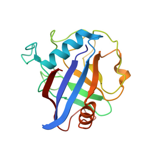
Structural and biochemical characterization of the cytosolic wheat cyclophilin TaCypA-1.

Sekhon, S.S.Kaur, H.Dutta, T.Singh, K.Kumari, S.Kang, S.Park, S.G.Park, B.C.Jeong, D.G.Pareek, A.Woo, E.J.Singh, P.Yoon, T.S.

(2013) Acta Crystallogr D Biol Crystallogr 69: 555-563

  • DOI: https://doi.org/10.1107/S0907444912051529
  • Primary Citation of Related Structures:  
    4E1Q, 4HY7

  • PubMed Abstract: 

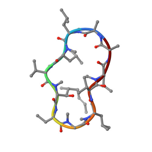
    Cyclophilins belong to a family of proteins that bind to the immunosuppressive drug cyclosporin A (CsA). Several members of this protein family catalyze the cis-trans isomerization of peptide bonds preceding prolyl residues. The present study describes the biochemical and structural characteristics of a cytosolic cyclophilin (TaCypA-1) cloned from wheat (Triticum aestivum L.). Purified TaCypA-1 expressed in Escherichia coli showed peptidyl-prolyl cis-trans isomerase activity, which was inhibited by CsA with an inhibition constant of 78.3 nM. The specific activity and catalytic efficiency (kcat/Km) of the purified TaCypA-1 were 99.06 ± 0.13 nmol s(-1) mg(-1) and 2.32 × 10(5) M(-1) s(-1), respectively. The structures of apo TaCypA-1 and the TaCypA-1-CsA complex were determined at 1.25 and 1.20 Å resolution, respectively, using X-ray diffraction. Binding of CsA to the active site of TaCypA-1 did not result in any significant conformational change in the apo TaCypA-1 structure. This is consistent with the crystal structure of the human cyclophilin D-CsA complex reported at 0.96 Å resolution. The TaCypA-1 structure revealed the presence of a divergent loop of seven amino acids (48)KSGKPLH(54) which is a characteristic feature of plant cyclophilins. This study is the first to elucidate the structure of an enzymatically active plant cyclophilin which shows peptidyl-prolyl cis-trans isomerase activity and the presence of a divergent loop.


  • Organizational Affiliation

    Medical Proteomics Research Center, Korea Research Institute of Bioscience and Biotechnology, Yuseong-gu, Daejeon, Republic of Korea.


Macromolecules
Find similar proteins by:  (by identity cutoff)  |  3D Structure
Entity ID: 1
MoleculeChains Sequence LengthOrganismDetailsImage
Peptidyl-prolyl cis-trans isomerase171Triticum aestivumMutation(s): 0 
Gene Names: CyP3CyP1
EC: 5.2.1.8
UniProt
Find proteins for Q93W25 (Triticum aestivum)
Explore Q93W25 
Go to UniProtKB:  Q93W25
Entity Groups  
Sequence Clusters30% Identity50% Identity70% Identity90% Identity95% Identity100% Identity
UniProt GroupQ93W25
Sequence Annotations
Expand
  • Reference Sequence

Find similar proteins by:  Sequence   |   3D Structure  

Entity ID: 2
MoleculeChains Sequence LengthOrganismDetailsImage
Cyclosporin A11Tolypocladium inflatumMutation(s): 0 
Entity Groups  
Sequence Clusters30% Identity50% Identity70% Identity90% Identity95% Identity100% Identity
Sequence Annotations
Expand
  • Reference Sequence
Small Molecules
Modified Residues  5 Unique
IDChains TypeFormula2D DiagramParent
ABA
Query on ABA
B
L-PEPTIDE LINKINGC4 H9 N O2ALA
BMT
Query on BMT
B
L-PEPTIDE LINKINGC10 H19 N O3THR
MLE
Query on MLE
B
L-PEPTIDE LINKINGC7 H15 N O2LEU
MVA
Query on MVA
B
L-PEPTIDE LINKINGC6 H13 N O2VAL
SAR
Query on SAR
B
PEPTIDE LINKINGC3 H7 N O2GLY
Biologically Interesting Molecules (External Reference) 1 Unique
Experimental Data & Validation

Experimental Data

  • Method: X-RAY DIFFRACTION
  • Resolution: 1.20 Å
  • R-Value Free: 0.204 
  • R-Value Work: 0.187 
  • R-Value Observed: 0.187 
  • Space Group: P 21 21 21
Unit Cell:
Length ( Å )Angle ( ˚ )
a = 37.914α = 90
b = 49.847β = 90
c = 78.976γ = 90
Software Package:
Software NamePurpose
HKL-2000data collection
PHENIXmodel building
REFMACrefinement
HKL-2000data reduction
HKL-2000data scaling
PHENIXphasing

Structure Validation

View Full Validation Report


Entry History 

Deposition Data

Revision History  (Full details and data files)

  • Version 1.0: 2013-03-27
    Type: Initial release
  • Version 1.1: 2014-09-17
    Changes: Database references
  • Version 1.2: 2023-11-08
    Changes: Data collection, Database references, Derived calculations, Refinement description
  • Version 1.3: 2023-12-06
    Changes: Data collection, Derived calculations