4HST | pdb_00004hst

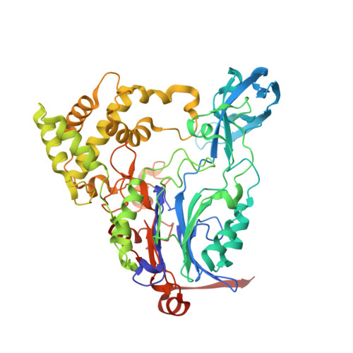
Crystal structure of a double mutant of a class III engineered cephalosporin acylase


Experimental Data Snapshot

  • Method: X-RAY DIFFRACTION
  • Resolution: 1.57 Å
  • R-Value Free: 
    0.162 (Depositor), 0.190 (DCC) 
  • R-Value Work: 
    0.119 (Depositor) 
  • R-Value Observed: 
    0.121 (Depositor) 

wwPDB Validation 3D Report Full Report

Validation slider image for 4HST

This is version 1.3 of the entry. See complete history. 

Literature

Structure of a class III engineered cephalosporin acylase: comparisons with class I acylase and implications for differences in substrate specificity and catalytic activity.

Golden, E., Paterson, R., Tie, W.J., Anandan, A., Flematti, G., Molla, G., Rosini, E., Pollegioni, L., Vrielink, A.

(2013) Biochem J 451: 217-226

  • DOI: https://doi.org/10.1042/BJ20121715
  • Primary Citation Related Structures: 
    4HSR, 4HST

  • PubMed Abstract: 

    The crystal structure of the wild-type form of glutaryl-7-ACA (7-aminocephalosporanic acid) acylase from Pseudomonas N176 and a double mutant of the protein (H57βS/H70βS) that displays enhanced catalytic efficiency on cephalosporin C over glutaryl-7-aminocephalosporanic acid has been determined. The structures show a heterodimer made up of an α-chain (229 residues) and a β-chain (543 residues) with a deep cavity, which constitutes the active site. Comparison of the wild-type and mutant structures provides insights into the molecular reasons for the observed enhanced specificity on cephalosporin C over glutaryl-7-aminocephalosporanic acid and offers the basis to evolve a further improved enzyme variant. The nucleophilic catalytic serine residue, Ser(1β), is situated at the base of the active site cavity. The electron density reveals a ligand covalently bound to the catalytic serine residue, such that a tetrahedral adduct is formed. This is proposed to mimic the transition state of the enzyme for both the maturation step and the catalysis of the substrates. A view of the transition state configuration of the enzyme provides important insights into the mechanism of substrate binding and catalysis.


  • Organizational Affiliation: 
    • School of Chemistry and Biochemistry, University of Western Australia, 35 Stirling Highway, Crawley, WA 6009, Australia.

Macromolecule Content 

  • Total Structure Weight: 84.23 kDa 
  • Atom Count: 6,863 
  • Modeled Residue Count: 751 
  • Deposited Residue Count: 772 
  • Unique protein chains: 2

Macromolecules

Find similar proteins by:|  3D Structure
Entity ID: 1
MoleculeChains  Sequence LengthOrganismDetailsImage
glutaryl-7-aminocephalosporanic acid acylase alpha chain229PseudomonasMutation(s): 0 
Entity Groups
Sequence Clusters30% Identity50% Identity70% Identity90% Identity95% Identity100% Identity
Sequence Annotations
Expand
Reference Sequence
Find similar proteins by:|  3D Structure
Entity ID: 2
MoleculeChains  Sequence LengthOrganismDetailsImage
glutaryl-7-aminocephalosporanic acid acylase beta chain543PseudomonasMutation(s): 0 
Entity Groups
Sequence Clusters30% Identity50% Identity70% Identity90% Identity95% Identity100% Identity
Sequence Annotations
Expand
Reference Sequence

Small Molecules

Ligands 1 Unique
IDChains Name / Formula / InChI Key2D Diagram3D Interactions
GLJ

Query on GLJ


Download:Ideal Coordinates CCD File
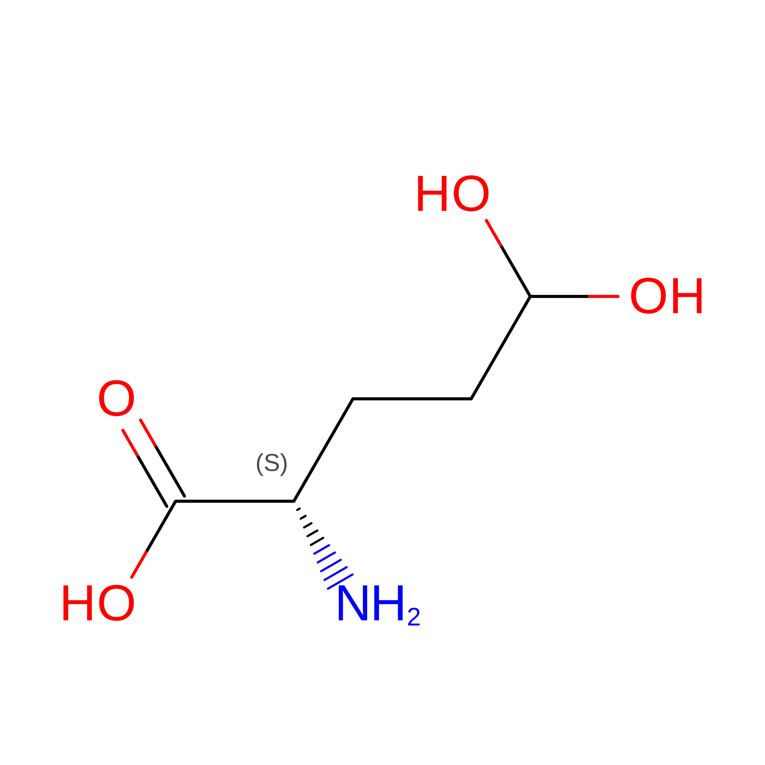
C [auth B]5,5-dihydroxy-L-norvaline
C5 H11 N O4
FIKXYBCGRJPSSD-VKHMYHEASA-N

Experimental Data & Validation

Experimental Data

  • Method: X-RAY DIFFRACTION
  • Resolution: 1.57 Å
  • R-Value Free:  0.162 (Depositor), 0.190 (DCC) 
  • R-Value Work:  0.119 (Depositor) 
  • R-Value Observed: 0.121 (Depositor) 
Space Group: P 21 21 21
Unit Cell:
Length ( Å )Angle ( ˚ )
a = 68.385α = 90
b = 77.845β = 90
c = 191.99γ = 90
Software Package:
Software NamePurpose
Blu-Icedata collection
SHELXSphasing
REFMACrefinement
MOSFLMdata reduction
SCALAdata scaling

Structure Validation

View Full Validation Report


Entry History 

Deposition Data

Revision History  (Full details and data files)

  • Version 1.0: 2013-02-27
    Type: Initial release
  • Version 1.1: 2013-04-10
    Changes: Database references
  • Version 1.2: 2017-11-15
    Changes: Refinement description
  • Version 1.3: 2024-02-28
    Changes: Data collection, Database references, Derived calculations