4HOV | pdb_00004hov

DypB N246A in complex with manganese


Experimental Data Snapshot

  • Method: X-RAY DIFFRACTION
  • Resolution: 2.20 Å
  • R-Value Free: 
    0.195 (Depositor), 0.190 (DCC) 
  • R-Value Work: 
    0.167 (Depositor) 
  • R-Value Observed: 
    0.169 (Depositor) 

Starting Model: experimental
View more details

wwPDB Validation 3D Report Full Report

Validation slider image for 4HOV

Ligand Structure Quality Assessment 


This is version 1.4 of the entry. See complete history

Literature

Improved Manganese-Oxidizing Activity of DypB, a Peroxidase from a Lignolytic Bacterium.

Singh, R.Grigg, J.C.Qin, W.Kadla, J.F.Murphy, M.E.Eltis, L.D.

(2013) ACS Chem Biol 8: 700-706

  • DOI: https://doi.org/10.1021/cb300608x
  • Primary Citation Related Structures: 
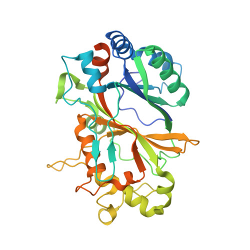
    4HOV

  • PubMed Abstract: 

    DypB, a dye-decolorizing peroxidase from the lignolytic soil bacterium Rhodococcus jostii RHA1, catalyzes the peroxide-dependent oxidation of divalent manganese (Mn(2+)), albeit less efficiently than fungal manganese peroxidases. Substitution of Asn246, a distal heme residue, with alanine increased the enzyme's apparent k(cat) and k(cat)/K(m) values for Mn(2+) by 80- and 15-fold, respectively. A 2.2 Å resolution X-ray crystal structure of the N246A variant revealed the Mn(2+) to be bound within a pocket of acidic residues at the heme edge, reminiscent of the binding site in fungal manganese peroxidase and very different from that of another bacterial Mn(2+)-oxidizing peroxidase. The first coordination sphere was entirely composed of solvent, consistent with the variant's high K(m) for Mn(2+) (17 ± 2 mM). N246A catalyzed the manganese-dependent transformation of hard wood kraft lignin and its solvent-extracted fractions. Two of the major degradation products were identified as 2,6-dimethoxybenzoquinone and 4-hydroxy-3,5-dimethoxybenzaldehyde, respectively. These results highlight the potential of bacterial enzymes as biocatalysts to transform lignin.


  • Organizational Affiliation
    • Department of Microbiology and Immunology, University of British Columbia, Vancouver, British Columbia, Canada.

Macromolecule Content 

  • Total Structure Weight: 114.74 kDa 
  • Atom Count: 8,014 
  • Modeled Residue Count: 925 
  • Deposited Residue Count: 1,059 
  • Unique protein chains: 1

Macromolecules

Find similar proteins by:|  3D Structure
Entity ID: 1
MoleculeChains  Sequence LengthOrganismDetailsImage
DypB
A, B, C
353Rhodococcus jostii RHA1Mutation(s): 1 
Gene Names: dypBRHA1_ro02407
EC: 1.11.1
UniProt
Find proteins for Q0SE24 (Rhodococcus jostii (strain RHA1))
Explore Q0SE24 
Go to UniProtKB:  Q0SE24
Entity Groups
Sequence Clusters30% Identity50% Identity70% Identity90% Identity95% Identity100% Identity
UniProt GroupQ0SE24
Sequence Annotations
Expand
Reference Sequence

Small Molecules

Ligands 5 Unique
IDChains Name / Formula / InChI Key2D Diagram3D Interactions
HEM

Query on HEM


Download:Ideal Coordinates CCD File
D [auth A],
H [auth B],
L [auth C]
PROTOPORPHYRIN IX CONTAINING FE
C34 H32 Fe N4 O4
KABFMIBPWCXCRK-RGGAHWMASA-L
GOL

Query on GOL


Download:Ideal Coordinates CCD File
K [auth B]GLYCEROL
C3 H8 O3
PEDCQBHIVMGVHV-UHFFFAOYSA-N
MN

Query on MN


Download:Ideal Coordinates CCD File
G [auth A],
J [auth B],
N [auth C]
MANGANESE (II) ION
Mn
WAEMQWOKJMHJLA-UHFFFAOYSA-N
FMT

Query on FMT


Download:Ideal Coordinates CCD File
F [auth A]FORMIC ACID
C H2 O2
BDAGIHXWWSANSR-UHFFFAOYSA-N
CL

Query on CL


Download:Ideal Coordinates CCD File
E [auth A],
I [auth B],
M [auth C]
CHLORIDE ION
Cl
VEXZGXHMUGYJMC-UHFFFAOYSA-M

Experimental Data & Validation

Experimental Data

  • Method: X-RAY DIFFRACTION
  • Resolution: 2.20 Å
  • R-Value Free:  0.195 (Depositor), 0.190 (DCC) 
  • R-Value Work:  0.167 (Depositor) 
  • R-Value Observed: 0.169 (Depositor) 
Space Group: P 32 2 1
Unit Cell:
Length ( Å )Angle ( ˚ )
a = 132.96α = 90
b = 132.96β = 90
c = 159.502γ = 120
Software Package:
Software NamePurpose
MOSFLMdata reduction
SCALAdata scaling
REFMACrefinement
PDB_EXTRACTdata extraction
Blu-Icedata collection
REFMACphasing

Structure Validation

View Full Validation Report


Ligand Structure Quality Assessment 


Entry History 

Deposition Data

Revision History  (Full details and data files)

  • Version 1.0: 2013-01-16
    Type: Initial release
  • Version 1.1: 2013-01-23
    Changes: Database references
  • Version 1.2: 2013-05-01
    Changes: Database references
  • Version 1.3: 2017-11-15
    Changes: Refinement description
  • Version 1.4: 2023-09-20
    Changes: Data collection, Database references, Derived calculations, Refinement description