4HN2 | pdb_00004hn2

Crystal structure of the catalytic domain of human diphosphoinositol pentakisphosphate kinase 2 (PPIP5K2) in complex with AMPPNP and a substrate analog 5PA-IP5


Experimental Data Snapshot

  • Method: X-RAY DIFFRACTION
  • Resolution: 1.90 Å
  • R-Value Free: 
    0.222 (Depositor), 0.219 (DCC) 
  • R-Value Work: 
    0.191 (Depositor), 0.190 (DCC) 
  • R-Value Observed: 
    0.193 (Depositor) 

Starting Model: experimental
View more details

wwPDB Validation 3D Report Full Report

Validation slider image for 4HN2

Ligand Structure Quality Assessment 


This is version 1.2 of the entry. See complete history

Literature

First synthetic analogues of diphosphoinositol polyphosphates: interaction with PP-InsP(5) kinase.

Riley, A.M.Wang, H.Weaver, J.D.Shears, S.B.Potter, B.V.

(2012) Chem Commun (Camb) 48: 11292-11294

  • DOI: https://doi.org/10.1039/c2cc36044f
  • Primary Citation Related Structures: 
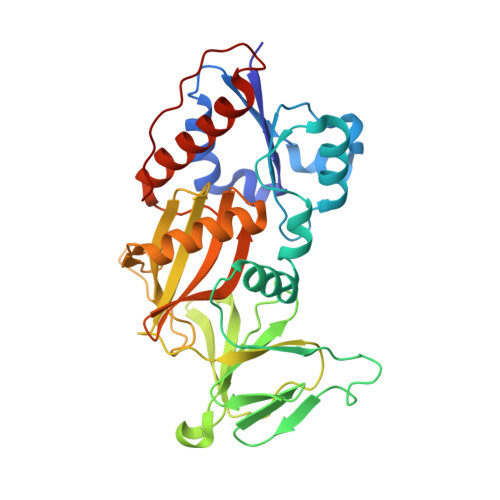
    4HN2

  • PubMed Abstract: 

    We synthesised analogues of diphosphoinositol polyphosphates (PP-InsPs) in which the diphosphate is replaced by an α-phosphonoacetic acid (PA) ester. Structural analysis revealed that 5-PA-InsP(5) mimics 5-PP-InsP(5) binding to the kinase domain of PPIP5K2; both molecules were phosphorylated by the enzyme. PA-InsPs are promising candidates for further studies into the biology of PP-InsPs.


  • Organizational Affiliation
    • Wolfson Laboratory of Medicinal Chemistry, Department of Pharmacy and Pharmacology, University of Bath, BA2 7AY, UK.

Macromolecule Content 

  • Total Structure Weight: 38.87 kDa 
  • Atom Count: 3,096 
  • Modeled Residue Count: 325 
  • Deposited Residue Count: 330 
  • Unique protein chains: 1

Macromolecules

Find similar proteins by:|  3D Structure
Entity ID: 1
MoleculeChains  Sequence LengthOrganismDetailsImage
Inositol hexakisphosphate and diphosphoinositol-pentakisphosphate kinase 2330Homo sapiensMutation(s): 0 
Gene Names: HISPPD1KIAA0433PPIP5K2VIP2
EC: 2.7.4.21 (PDB Primary Data), 2.7.4.24 (PDB Primary Data)
UniProt & NIH Common Fund Data Resources
Find proteins for O43314 (Homo sapiens)
Explore O43314 
Go to UniProtKB:  O43314
PHAROS:  O43314
GTEx:  ENSG00000145725 
Entity Groups
Sequence Clusters30% Identity50% Identity70% Identity90% Identity95% Identity100% Identity
UniProt GroupO43314
Sequence Annotations
Expand
Reference Sequence

Small Molecules

Ligands 3 Unique
IDChains Name / Formula / InChI Key2D Diagram3D Interactions
0EJ

Query on 0EJ


Download:Ideal Coordinates CCD File
C [auth A](2-oxo-2-{[(1s,2R,3S,4s,5R,6S)-2,3,4,5,6-pentakis(phosphonooxy)cyclohexyl]oxy}ethyl)phosphonic acid
C8 H20 O25 P6
JGWVNODNEALCIO-QEEWUKHFSA-N
ANP

Query on ANP


Download:Ideal Coordinates CCD File
B [auth A]PHOSPHOAMINOPHOSPHONIC ACID-ADENYLATE ESTER
C10 H17 N6 O12 P3
PVKSNHVPLWYQGJ-KQYNXXCUSA-N
MG

Query on MG


Download:Ideal Coordinates CCD File
D [auth A],
E [auth A],
F [auth A],
G [auth A]
MAGNESIUM ION
Mg
JLVVSXFLKOJNIY-UHFFFAOYSA-N

Experimental Data & Validation

Experimental Data

  • Method: X-RAY DIFFRACTION
  • Resolution: 1.90 Å
  • R-Value Free:  0.222 (Depositor), 0.219 (DCC) 
  • R-Value Work:  0.191 (Depositor), 0.190 (DCC) 
  • R-Value Observed: 0.193 (Depositor) 
Space Group: P 21 21 21
Unit Cell:
Length ( Å )Angle ( ˚ )
a = 89.666α = 90
b = 110.768β = 90
c = 41.536γ = 90
Software Package:
Software NamePurpose
HKL-2000data collection
REFMACrefinement
HKL-2000data reduction
HKL-2000data scaling
REFMACphasing

Structure Validation

View Full Validation Report


Ligand Structure Quality Assessment 


Entry History 

Deposition Data

  • Released Date: 2012-10-31 
  • Deposition Author(s): Wang, H.
  • This entry supersedes: 4GB4

Revision History  (Full details and data files)

  • Version 1.0: 2012-10-31
    Type: Initial release
  • Version 1.1: 2012-11-07
    Changes: Database references
  • Version 1.2: 2023-09-20
    Changes: Data collection, Database references, Derived calculations, Refinement description