4HJV

Crystal structure of E. coli MltE with bound bulgecin and murodipeptide


Experimental Data Snapshot

  • Method: X-RAY DIFFRACTION
  • Resolution: 2.30 Å
  • R-Value Free: 0.203 
  • R-Value Work: 0.163 
  • R-Value Observed: 0.167 

wwPDB Validation   3D Report Full Report


Ligand Structure Quality Assessment 


This is version 2.1 of the entry. See complete history


Literature

On the Mechanism of Peptidoglycan Binding and Cleavage by the endo-Specific Lytic Transglycosylase MltE from Escherichia coli.

Fibriansah, G.Gliubich, F.I.Thunnissen, A.M.

(2012) Biochemistry 51: 9164-9177

  • DOI: https://doi.org/10.1021/bi300900t
  • Primary Citation of Related Structures:  
    3T36, 4HJV, 4HJY, 4HJZ

  • PubMed Abstract: 

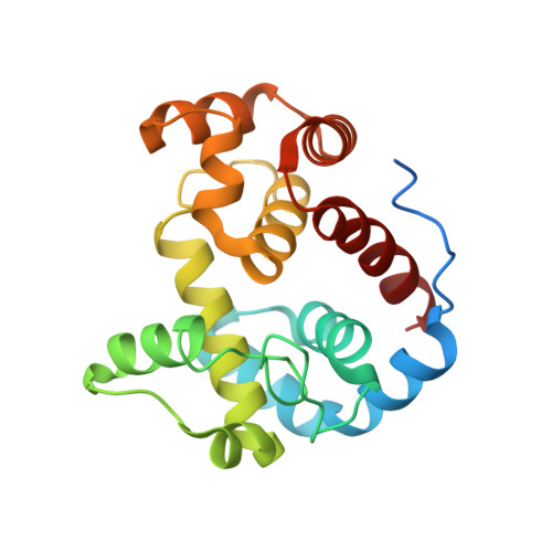
    The lytic transglycosylase MltE from Escherichia coli is a periplasmic, outer membrane-attached enzyme that cleaves the β-1,4-glycosidic bonds between N-acetylmuramic acid and N-acetylglucosamine residues in the cell wall peptidoglycan, producing 1,6-anhydromuropeptides. Here we report three crystal structures of MltE: in a substrate-free state, in a binary complex with chitopentaose, and in a ternary complex with the glycopeptide inhibitor bulgecin A and the murodipeptide N-acetylglucosaminyl-N-acetylmuramyl-l-Ala-d-Glu. The substrate-bound structures allowed a detailed analysis of the saccharide-binding interactions in six subsites of the peptidoglycan-binding groove (subsites -4 to +2) and, combined with site-directed mutagenesis analysis, confirmed the role of Glu64 as catalytic acid/base. The structures permitted the precise modeling of a short glycan strand of eight saccharide residues, providing evidence for two additional subsites (+3 and +4) and revealing the productive conformational state of the substrate at subsites -1 and +1, where the glycosidic bond is cleaved. Full accessibility of the peptidoglycan-binding groove and preferential binding of an N-acetylmuramic acid residue in a (4)C(1) chair conformation at subsite +2 explain why MltE shows only endo- and no exo-specific activity toward glycan strands. The results further indicate that catalysis of glycosidic bond cleavage by MltE proceeds via distortion toward a sofa-like conformation of the N-acetylmuramic acid sugar ring at subsite -1 and by anchimeric assistance of the sugar's N-acetyl group, as shown previously for the lytic transglycosylases Slt70 and MltB.


  • Organizational Affiliation

    Laboratory of Biophysical Chemistry, Groningen Biomolecular Sciences and Biotechnology Institute, University of Groningen, Nijenborgh 7, 9747 AG Groningen, The Netherlands.


Macromolecules
Find similar proteins by:  (by identity cutoff)  |  3D Structure
Entity ID: 1
MoleculeChains Sequence LengthOrganismDetailsImage
Endo-type membrane-bound lytic murein transglycosylase A
A, B, C, D, E
203Escherichia coli K-12Mutation(s): 0 
Gene Names: emtAmltEsltZycgPb1193JW5821
EC: 4.2.2
UniProt
Find proteins for P0C960 (Escherichia coli (strain K12))
Explore P0C960 
Go to UniProtKB:  P0C960
Entity Groups  
Sequence Clusters30% Identity50% Identity70% Identity90% Identity95% Identity100% Identity
UniProt GroupP0C960
Sequence Annotations
Expand
  • Reference Sequence
Oligosaccharides

Help

Entity ID: 2
MoleculeChains Length2D Diagram Glycosylation3D Interactions
2-acetamido-2-deoxy-beta-D-glucopyranose-(1-4)-N-acetyl-beta-muramic acid
F, G, H
2N/A
Glycosylation Resources
GlyTouCan:  G99515FY
GlyCosmos:  G99515FY
Experimental Data & Validation

Experimental Data

  • Method: X-RAY DIFFRACTION
  • Resolution: 2.30 Å
  • R-Value Free: 0.203 
  • R-Value Work: 0.163 
  • R-Value Observed: 0.167 
  • Space Group: P 21 21 21
Unit Cell:
Length ( Å )Angle ( ˚ )
a = 78.778α = 90
b = 94.853β = 90
c = 162.016γ = 90
Software Package:
Software NamePurpose
MAR345dtbdata collection
MOLREPphasing
PHENIXrefinement
DENZOdata reduction
SCALEPACKdata scaling
SCALAdata scaling

Structure Validation

View Full Validation Report


Ligand Structure Quality Assessment 


Entry History 

Deposition Data

Revision History  (Full details and data files)

  • Version 1.0: 2012-10-24
    Type: Initial release
  • Version 1.1: 2012-10-31
    Changes: Database references
  • Version 1.2: 2012-12-26
    Changes: Database references
  • Version 1.3: 2017-11-15
    Changes: Refinement description
  • Version 2.0: 2020-07-29
    Type: Remediation
    Reason: Carbohydrate remediation
    Changes: Atomic model, Data collection, Database references, Derived calculations, Structure summary
  • Version 2.1: 2023-09-20
    Changes: Data collection, Database references, Refinement description, Structure summary