4HFZ

Crystal Structure of an MDM2/P53 Peptide Complex


Experimental Data Snapshot

  • Method: X-RAY DIFFRACTION
  • Resolution: 2.69 Å
  • R-Value Free: 0.247 
  • R-Value Work: 0.195 
  • R-Value Observed: 0.198 

wwPDB Validation   3D Report Full Report


This is version 1.2 of the entry. See complete history


Literature

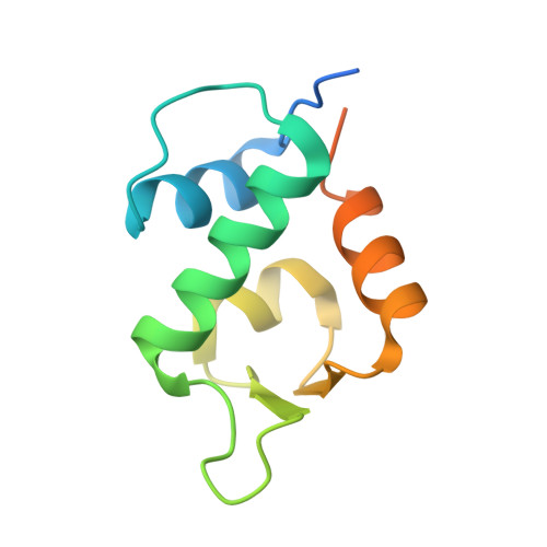
The structure of an MDM2-Nutlin-3a complex solved by the use of a validated MDM2 surface-entropy reduction mutant.

Anil, B.Riedinger, C.Endicott, J.A.Noble, M.E.

(2013) Acta Crystallogr D Biol Crystallogr 69: 1358-1366

  • DOI: https://doi.org/10.1107/S0907444913004459
  • Primary Citation of Related Structures:  
    4HFZ, 4HG7

  • PubMed Abstract: 

    The p53-binding site of MDM2 holds great promise as a target for therapeutic intervention in MDM2-amplified p53 wild-type forms of cancer. Despite the extensive validation of this strategy, there are relatively few crystallographically determined co-complex structures for small-molecular inhibitors of the MDM2-p53 interaction available in the PDB. Here, a surface-entropy reduction mutant of the N-terminal domain of MDM2 that has been designed to enhance crystallogenesis is presented. This mutant has been validated by comparative ligand-binding studies using differential scanning fluorimetry and fluorescence polarization anisotropy and by cocrystallization with a peptide derived from p53. Using this mutant, the cocrystal structure of MDM2 with the benchmark inhibitor Nutlin-3a has been determined, revealing subtle differences from the previously described co-complex of MDM2 with Nutlin-2.


  • Organizational Affiliation

    Department of Biochemistry, University of Oxford, South Parks Road, Oxford OX1 3QU, England.


Macromolecules
Find similar proteins by:  (by identity cutoff)  |  3D Structure
Entity ID: 1
MoleculeChains Sequence LengthOrganismDetailsImage
E3 ubiquitin-protein ligase Mdm2
A, C
109Homo sapiensMutation(s): 2 
Gene Names: MDM2
EC: 6.3.2
UniProt & NIH Common Fund Data Resources
Find proteins for Q00987 (Homo sapiens)
Explore Q00987 
Go to UniProtKB:  Q00987
PHAROS:  Q00987
GTEx:  ENSG00000135679 
Entity Groups  
Sequence Clusters30% Identity50% Identity70% Identity90% Identity95% Identity100% Identity
UniProt GroupQ00987
Sequence Annotations
Expand
  • Reference Sequence

Find similar proteins by:  Sequence   |   3D Structure  

Entity ID: 2
MoleculeChains Sequence LengthOrganismDetailsImage
Cellular tumor antigen p53
B, D
15Homo sapiensMutation(s): 0 
UniProt & NIH Common Fund Data Resources
Find proteins for P04637 (Homo sapiens)
Explore P04637 
Go to UniProtKB:  P04637
PHAROS:  P04637
GTEx:  ENSG00000141510 
Entity Groups  
Sequence Clusters30% Identity50% Identity70% Identity90% Identity95% Identity100% Identity
UniProt GroupP04637
Sequence Annotations
Expand
  • Reference Sequence
Small Molecules
Ligands 1 Unique
IDChains Name / Formula / InChI Key2D Diagram3D Interactions
SO4
Query on SO4

Download Ideal Coordinates CCD File 
E [auth A]SULFATE ION
O4 S
QAOWNCQODCNURD-UHFFFAOYSA-L
Experimental Data & Validation

Experimental Data

  • Method: X-RAY DIFFRACTION
  • Resolution: 2.69 Å
  • R-Value Free: 0.247 
  • R-Value Work: 0.195 
  • R-Value Observed: 0.198 
  • Space Group: P 43 21 2
Unit Cell:
Length ( Å )Angle ( ˚ )
a = 52.944α = 90
b = 52.944β = 90
c = 196.201γ = 90
Software Package:
Software NamePurpose
XDSdata scaling
PHASERphasing
REFMACrefinement
XDSdata reduction
SCALAdata scaling

Structure Validation

View Full Validation Report


Entry History 

Deposition Data

Revision History  (Full details and data files)

  • Version 1.0: 2013-07-31
    Type: Initial release
  • Version 1.1: 2013-08-21
    Changes: Database references
  • Version 1.2: 2024-02-28
    Changes: Data collection, Database references, Derived calculations, Refinement description