4HE5 | pdb_00004he5

Crystal structure of the selenomethionine variant of the C-terminal domain of Geobacillus thermoleovorans putative U32 peptidase


Experimental Data Snapshot

  • Method: X-RAY DIFFRACTION
  • Resolution: 1.15 Å
  • R-Value Free: 
    0.170 (Depositor), 0.174 (DCC) 
  • R-Value Work: 
    0.141 (Depositor), 0.137 (DCC) 
  • R-Value Observed: 
    0.141 (Depositor) 

wwPDB Validation 3D Report Full Report

Validation slider image for 4HE5

This is version 1.5 of the entry. See complete history

Literature

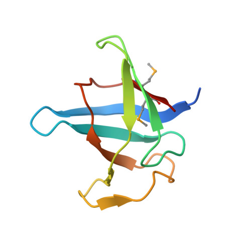
Ultratight crystal packing of a 10 kDa protein.

Trillo-Muyo, S.Jasilionis, A.Domagalski, M.J.Chruszcz, M.Minor, W.Kuisiene, N.Arolas, J.L.Sola, M.Gomis-Ruth, F.X.

(2013) Acta Crystallogr D Biol Crystallogr 69: 464-470

  • DOI: https://doi.org/10.1107/S0907444912050135
  • Primary Citation Related Structures: 
    4HE5, 4HE6

  • PubMed Abstract: 

    While small organic molecules generally crystallize forming tightly packed lattices with little solvent content, proteins form air-sensitive high-solvent-content crystals. Here, the crystallization and full structure analysis of a novel recombinant 10 kDa protein corresponding to the C-terminal domain of a putative U32 peptidase are reported. The orthorhombic crystal contained only 24.5% solvent and is therefore among the most tightly packed protein lattices ever reported.


  • Organizational Affiliation
    • Proteolysis Laboratory, Department of Structural Biology, Molecular Biology Institute of Barcelona, Spanish Research Council CSIC, Barcelona Science Park, c/Baldiri Reixac 15-21, 08028 Barcelona, Spain.

Macromolecule Content 

  • Total Structure Weight: 10.6 kDa 
  • Atom Count: 777 
  • Modeled Residue Count: 82 
  • Deposited Residue Count: 89 
  • Unique protein chains: 1

Macromolecules

Find similar proteins by:|  3D Structure
Entity ID: 1
MoleculeChains  Sequence LengthOrganismDetailsImage
Peptidase family U3289Geobacillus thermoleovorans CCB_US3_UF5Mutation(s): 0 

Small Molecules

Ligands 1 Unique
IDChains Name / Formula / InChI Key2D Diagram3D Interactions
SO4

Query on SO4


Download:Ideal Coordinates CCD File
B [auth A]SULFATE ION
O4 S
QAOWNCQODCNURD-UHFFFAOYSA-L
Modified Residues  1 Unique
IDChains TypeFormula2D DiagramParent
MSE
Query on MSE
A
L-PEPTIDE LINKINGC5 H11 N O2 SeMET

Experimental Data & Validation

Experimental Data

  • Method: X-RAY DIFFRACTION
  • Resolution: 1.15 Å
  • R-Value Free:  0.170 (Depositor), 0.174 (DCC) 
  • R-Value Work:  0.141 (Depositor), 0.137 (DCC) 
  • R-Value Observed: 0.141 (Depositor) 
Space Group: P 32 2 1
Unit Cell:
Length ( Å )Angle ( ˚ )
a = 45.55α = 90
b = 45.55β = 90
c = 60.9γ = 120
Software Package:
Software NamePurpose
ProDCdata collection
SHELXmodel building
BUSTERrefinement
XDSdata reduction
SCALAdata scaling
SHELXphasing

Structure Validation

View Full Validation Report


Entry History 

Deposition Data

Revision History  (Full details and data files)

  • Version 1.0: 2012-11-14
    Type: Initial release
  • Version 1.1: 2013-02-27
    Changes: Database references
  • Version 1.2: 2013-06-19
    Changes: Database references
  • Version 1.3: 2017-11-15
    Changes: Refinement description
  • Version 1.4: 2022-04-13
    Changes: Database references, Derived calculations, Structure summary
  • Version 1.5: 2024-11-06
    Changes: Data collection, Structure summary