4HCZ

PHF1 Tudor in complex with H3K36me3


Experimental Data Snapshot

  • Method: X-RAY DIFFRACTION
  • Resolution: 1.85 Å
  • R-Value Free: 0.244 
  • R-Value Work: 0.204 
  • R-Value Observed: 0.206 

wwPDB Validation   3D Report Full Report


This is version 1.2 of the entry. See complete history


Literature

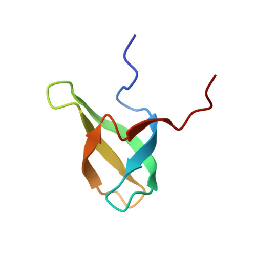
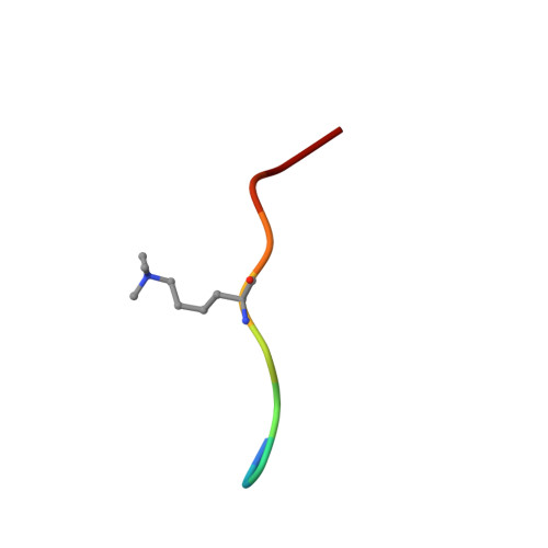
Molecular basis for H3K36me3 recognition by the Tudor domain of PHF1.

Musselman, C.A.Avvakumov, N.Watanabe, R.Abraham, C.G.Lalonde, M.E.Hong, Z.Allen, C.Roy, S.Nunez, J.K.Nickoloff, J.Kulesza, C.A.Yasui, A.Cote, J.Kutateladze, T.G.

(2012) Nat Struct Mol Biol 19: 1266-1272

  • DOI: https://doi.org/10.1038/nsmb.2435
  • Primary Citation of Related Structures:  
    4HCZ

  • PubMed Abstract: 

    The PHD finger protein 1 (PHF1) is essential in epigenetic regulation and genome maintenance. Here we show that the Tudor domain of human PHF1 binds to histone H3 trimethylated at Lys36 (H3K36me3). We report a 1.9-Å resolution crystal structure of the Tudor domain in complex with H3K36me3 and describe the molecular mechanism of H3K36me3 recognition using NMR. Binding of PHF1 to H3K36me3 inhibits the ability of the Polycomb PRC2 complex to methylate Lys27 of histone H3 in vitro and in vivo. Laser microirradiation data show that PHF1 is transiently recruited to DNA double-strand breaks, and PHF1 mutants impaired in the H3K36me3 interaction exhibit reduced retention at double-strand break sites. Together, our findings suggest that PHF1 can mediate deposition of the repressive H3K27me3 mark and acts as a cofactor in early DNA-damage response.


  • Organizational Affiliation

    Department of Pharmacology, University of Colorado School of Medicine, Aurora, Colorado, USA.


Macromolecules
Find similar proteins by:  (by identity cutoff)  |  3D Structure
Entity ID: 1
MoleculeChains Sequence LengthOrganismDetailsImage
PHD finger protein 1
A, B
58Homo sapiensMutation(s): 0 
Gene Names: PHF1PCL1
UniProt & NIH Common Fund Data Resources
Find proteins for O43189 (Homo sapiens)
Explore O43189 
Go to UniProtKB:  O43189
PHAROS:  O43189
GTEx:  ENSG00000112511 
Entity Groups  
Sequence Clusters30% Identity50% Identity70% Identity90% Identity95% Identity100% Identity
UniProt GroupO43189
Sequence Annotations
Expand
  • Reference Sequence

Find similar proteins by:  Sequence   |   3D Structure  

Entity ID: 2
MoleculeChains Sequence LengthOrganismDetailsImage
H3L-like histone
C, D
10Homo sapiensMutation(s): 0 
UniProt
Find proteins for Q6TXQ4 (Homo sapiens)
Explore Q6TXQ4 
Go to UniProtKB:  Q6TXQ4
Entity Groups  
Sequence Clusters30% Identity50% Identity70% Identity90% Identity95% Identity100% Identity
UniProt GroupQ6TXQ4
Sequence Annotations
Expand
  • Reference Sequence
Small Molecules
Modified Residues  1 Unique
IDChains TypeFormula2D DiagramParent
M3L
Query on M3L
C, D
L-PEPTIDE LINKINGC9 H21 N2 O2LYS
Experimental Data & Validation

Experimental Data

  • Method: X-RAY DIFFRACTION
  • Resolution: 1.85 Å
  • R-Value Free: 0.244 
  • R-Value Work: 0.204 
  • R-Value Observed: 0.206 
  • Space Group: C 1 2 1
Unit Cell:
Length ( Å )Angle ( ˚ )
a = 88.03α = 90
b = 29.67β = 116.41
c = 62γ = 90
Software Package:
Software NamePurpose
PHASERphasing
PHENIXrefinement
PDB_EXTRACTdata extraction
CrystalCleardata collection
d*TREKdata reduction
d*TREKdata scaling

Structure Validation

View Full Validation Report


Entry History 

Deposition Data

Revision History  (Full details and data files)

  • Version 1.0: 2012-11-14
    Type: Initial release
  • Version 1.1: 2012-12-19
    Changes: Database references
  • Version 1.2: 2023-09-20
    Changes: Data collection, Database references, Derived calculations, Refinement description