4H4O | pdb_00004h4o

Crystal Structure of HIV-1 Reverse Transcriptase (RT) in Complex with (E)-3-(3-(2-(2-(2,4-dioxo-3,4-dihydropyrimidin-1(2H)-yl)ethoxy)- 4-fluorophenoxy)-5-fluorophenyl)acrylonitrile (JLJ506), A Non-nucleoside inhibitor


Experimental Data Snapshot

  • Method: X-RAY DIFFRACTION
  • Resolution: 2.90 Å
  • R-Value Free: 
    0.265 (Depositor), 0.263 (DCC) 
  • R-Value Work: 
    0.232 (Depositor), 0.233 (DCC) 
  • R-Value Observed: 
    0.232 (Depositor) 

Starting Model: experimental
View more details

wwPDB Validation 3D Report Full Report

Validation slider image for 4H4O

Ligand Structure Quality Assessment 


This is version 1.3 of the entry. See complete history

Literature

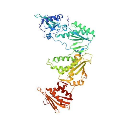
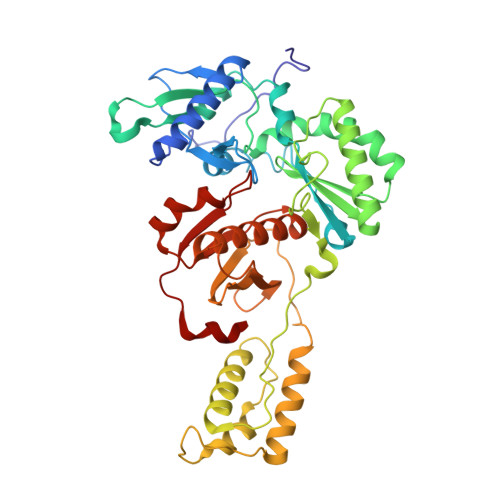
Crystal Structures of HIV-1 Reverse Transcriptase with Picomolar Inhibitors Reveal Key Interactions for Drug Design.

Frey, K.M.Bollini, M.Mislak, A.C.Cisneros, J.A.Gallardo-Macias, R.Jorgensen, W.L.Anderson, K.S.

(2012) J Am Chem Soc 134: 19501-19503

  • DOI: https://doi.org/10.1021/ja3092642
  • Primary Citation Related Structures: 
    4H4M, 4H4O

  • PubMed Abstract: 

    X-ray crystal structures at 2.9 Å resolution are reported for two complexes of catechol diethers with HIV-1 reverse transcriptase. The results help elucidate the structural origins of the extreme antiviral activity of the compounds. The possibility of halogen bonding between the inhibitors and Pro95 is addressed. Structural analysis reveals key interactions with conserved residues P95 and W229 of importance for design of inhibitors with high potency and favorable resistance profiles.


  • Organizational Affiliation
    • Department of Chemistry, Yale University, New Haven, Connecticut 06520-8107, United States.

Macromolecule Content 

  • Total Structure Weight: 114.44 kDa 
  • Atom Count: 8,082 
  • Modeled Residue Count: 977 
  • Deposited Residue Count: 985 
  • Unique protein chains: 2

Macromolecules

Find similar proteins by:|  3D Structure
Entity ID: 1
MoleculeChains  Sequence LengthOrganismDetailsImage
Reverse transcriptase/ribonuclease H, Exoribonuclease H, p66 RT557Human immunodeficiency virus type 1 BH10Mutation(s): 3 
Gene Names: p66 RT
EC: 2.7.7.49 (PDB Primary Data), 2.7.7.7 (PDB Primary Data), 3.1.26.13 (PDB Primary Data), 3.1.13.2 (PDB Primary Data)
UniProt
Find proteins for P03366 (Human immunodeficiency virus type 1 group M subtype B (isolate BH10))
Explore P03366 
Go to UniProtKB:  P03366
Entity Groups
Sequence Clusters30% Identity50% Identity70% Identity90% Identity95% Identity100% Identity
UniProt GroupP03366
Sequence Annotations
Expand
Reference Sequence
Find similar proteins by:|  3D Structure
Entity ID: 2
MoleculeChains  Sequence LengthOrganismDetailsImage
Reverse transcriptase/ribonuclease H, Exoribonuclease H, p51 RT428Human immunodeficiency virus type 1 BH10Mutation(s): 1 
Gene Names: p51 RT
EC: 2.7.7.49 (PDB Primary Data), 2.7.7.7 (PDB Primary Data), 3.1.26.13 (PDB Primary Data), 3.1.13.2 (PDB Primary Data)
UniProt
Find proteins for P03366 (Human immunodeficiency virus type 1 group M subtype B (isolate BH10))
Explore P03366 
Go to UniProtKB:  P03366
Entity Groups
Sequence Clusters30% Identity50% Identity70% Identity90% Identity95% Identity100% Identity
UniProt GroupP03366
Sequence Annotations
Expand
Reference Sequence

Small Molecules

Ligands 1 Unique
IDChains Name / Formula / InChI Key2D Diagram3D Interactions
506

Query on 506


Download:Ideal Coordinates CCD File
C [auth A](2E)-3-(3-{2-[2-(2,4-dioxo-3,4-dihydropyrimidin-1(2H)-yl)ethoxy]-4-fluorophenoxy}-5-fluorophenyl)prop-2-enenitrile
C21 H15 F2 N3 O4
USRHSVZSPKDGMT-OWOJBTEDSA-N
Binding Affinity Annotations 
IDSourceBinding Affinity
506 BindingDB:  4H4O IC50: 2.7 (nM) from 1 assay(s)

Experimental Data & Validation

Experimental Data

  • Method: X-RAY DIFFRACTION
  • Resolution: 2.90 Å
  • R-Value Free:  0.265 (Depositor), 0.263 (DCC) 
  • R-Value Work:  0.232 (Depositor), 0.233 (DCC) 
  • R-Value Observed: 0.232 (Depositor) 
Space Group: C 1 2 1
Unit Cell:
Length ( Å )Angle ( ˚ )
a = 225.451α = 90
b = 69.575β = 106.21
c = 104.401γ = 90
Software Package:
Software NamePurpose
DENZOdata reduction
SCALEPACKdata scaling
PHENIXrefinement
PDB_EXTRACTdata extraction
CBASSdata collection
HKL-2000data reduction
HKL-2000data scaling
PHASERphasing

Structure Validation

View Full Validation Report


Ligand Structure Quality Assessment 


Entry History 

Deposition Data

Revision History  (Full details and data files)

  • Version 1.0: 2012-12-05
    Type: Initial release
  • Version 1.1: 2013-01-02
    Changes: Database references
  • Version 1.2: 2017-11-15
    Changes: Refinement description
  • Version 1.3: 2023-09-20
    Changes: Data collection, Database references, Derived calculations, Refinement description