4GZ8

Mouse Semaphorin 3A, domains Sema-PSI-IG


Experimental Data Snapshot

  • Method: X-RAY DIFFRACTION
  • Resolution: 3.30 Å
  • R-Value Free: 0.265 
  • R-Value Work: 0.213 
  • R-Value Observed: 0.216 

wwPDB Validation   3D Report Full Report


Ligand Structure Quality Assessment 


This is version 2.1 of the entry. See complete history


Literature

Neuropilins lock secreted semaphorins onto plexins in a ternary signaling complex.

Janssen, B.J.C.Malinauskas, T.Weir, G.A.Cader, M.Z.Siebold, C.Jones, E.Y.

(2012) Nat Struct Mol Biol 19: 1293-1299

  • DOI: https://doi.org/10.1038/nsmb.2416
  • Primary Citation of Related Structures:  
    4GZ8, 4GZ9, 4GZA

  • PubMed Abstract: 

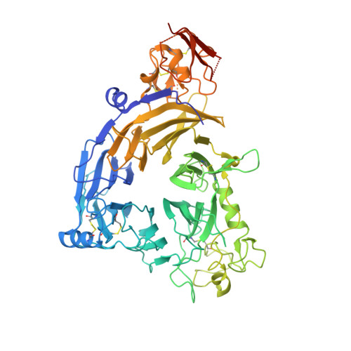
    Co-receptors add complexity to cell-cell signaling systems. The secreted semaphorin 3s (Sema3s) require a co-receptor, neuropilin (Nrp), to signal through plexin As (PlxnAs) in functions ranging from axon guidance to bone homeostasis, but the role of the co-receptor is obscure. Here we present the low-resolution crystal structure of a mouse semaphorin-plexin-Nrp complex alongside unliganded component structures. Dimeric semaphorin, two copies of plexin and two copies of Nrp are arranged as a dimer of heterotrimers. In each heterotrimer subcomplex, semaphorin contacts plexin, similar to in co-receptor-independent signaling complexes. The Nrp1s cross brace the assembly, bridging between sema domains of the Sema3A and PlxnA2 subunits from the two heterotrimers. Biophysical and cellular analyses confirm that this Nrp binding mode stabilizes a canonical, but weakened, Sema3-PlxnA interaction, adding co-receptor control over the mechanism by which receptor dimerization and/or oligomerization triggers signaling.


  • Organizational Affiliation

    Division of Structural Biology, Wellcome Trust Centre for Human Genetics, University of Oxford, Oxford, UK.


Macromolecules
Find similar proteins by:  (by identity cutoff)  |  3D Structure
Entity ID: 1
MoleculeChains Sequence LengthOrganismDetailsImage
Semaphorin-3A
A, B
667Mus musculusMutation(s): 2 
Gene Names: Sema3aSemadSemD
UniProt
Find proteins for O08665 (Mus musculus)
Explore O08665 
Go to UniProtKB:  O08665
Entity Groups  
Sequence Clusters30% Identity50% Identity70% Identity90% Identity95% Identity100% Identity
UniProt GroupO08665
Sequence Annotations
Expand
  • Reference Sequence
Oligosaccharides

Help

Entity ID: 2
MoleculeChains Length2D Diagram Glycosylation3D Interactions
alpha-D-mannopyranose-(1-3)-beta-D-mannopyranose-(1-4)-2-acetamido-2-deoxy-beta-D-glucopyranose-(1-4)-2-acetamido-2-deoxy-beta-D-glucopyranose
C
4N-Glycosylation
Glycosylation Resources
GlyTouCan:  G81315DD
GlyCosmos:  G81315DD
GlyGen:  G81315DD
Entity ID: 3
MoleculeChains Length2D Diagram Glycosylation3D Interactions
2-acetamido-2-deoxy-beta-D-glucopyranose-(1-4)-2-acetamido-2-deoxy-beta-D-glucopyranose
D
2N-Glycosylation
Glycosylation Resources
GlyTouCan:  G42666HT
GlyCosmos:  G42666HT
GlyGen:  G42666HT
Entity ID: 4
MoleculeChains Length2D Diagram Glycosylation3D Interactions
alpha-D-mannopyranose-(1-6)-beta-D-mannopyranose-(1-4)-2-acetamido-2-deoxy-beta-D-glucopyranose-(1-4)-2-acetamido-2-deoxy-beta-D-glucopyranose
E
4N-Glycosylation
Glycosylation Resources
GlyTouCan:  G22573RC
GlyCosmos:  G22573RC
GlyGen:  G22573RC
Small Molecules
Ligands 3 Unique
IDChains Name / Formula / InChI Key2D Diagram3D Interactions
NAG
Query on NAG

Download Ideal Coordinates CCD File 
L [auth B]2-acetamido-2-deoxy-beta-D-glucopyranose
C8 H15 N O6
OVRNDRQMDRJTHS-FMDGEEDCSA-N
ACT
Query on ACT

Download Ideal Coordinates CCD File 
I [auth A],
M [auth B]
ACETATE ION
C2 H3 O2
QTBSBXVTEAMEQO-UHFFFAOYSA-M
CA
Query on CA

Download Ideal Coordinates CCD File 
F [auth A],
G [auth A],
H [auth A],
J [auth B],
K [auth B]
CALCIUM ION
Ca
BHPQYMZQTOCNFJ-UHFFFAOYSA-N
Experimental Data & Validation

Experimental Data

  • Method: X-RAY DIFFRACTION
  • Resolution: 3.30 Å
  • R-Value Free: 0.265 
  • R-Value Work: 0.213 
  • R-Value Observed: 0.216 
  • Space Group: P 21 21 21
Unit Cell:
Length ( Å )Angle ( ˚ )
a = 97.63α = 90
b = 126.17β = 90
c = 161.48γ = 90
Software Package:
Software NamePurpose
GDAdata collection
PHASERphasing
REFMACrefinement
MOSFLMdata reduction
Aimlessdata scaling

Structure Validation

View Full Validation Report


Ligand Structure Quality Assessment 


Entry History 

Deposition Data

Revision History  (Full details and data files)

  • Version 1.0: 2012-10-17
    Type: Initial release
  • Version 1.1: 2012-11-07
    Changes: Database references
  • Version 1.2: 2012-12-26
    Changes: Database references
  • Version 2.0: 2020-07-29
    Type: Remediation
    Reason: Carbohydrate remediation
    Changes: Atomic model, Data collection, Database references, Derived calculations, Structure summary
  • Version 2.1: 2023-11-08
    Changes: Data collection, Database references, Refinement description, Structure summary