


Lysine Carboxylation: Metal and Structure Requirements for Post-translational Modification
Hsien, Y.C., Chen, M.C., Hsu, C.C., Chan, S.I., Yang, Y.S., Chen, C.J.To be published.
Experimental Data Snapshot
Entity ID: 1 | |||||
|---|---|---|---|---|---|
| Molecule | Chains | Sequence Length | Organism | Details | Image |
| dihydropyrimidinase | 500 | Tetraodon nigroviridis | Mutation(s): 0 Gene Names: DPYS | 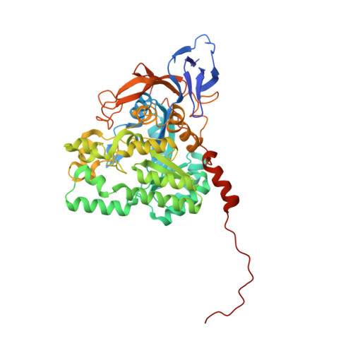 | |
UniProt | |||||
Entity Groups | |||||
| Sequence Clusters | 30% Identity50% Identity70% Identity90% Identity95% Identity100% Identity | ||||
| UniProt Group | H3C542 | ||||
Sequence AnnotationsExpand | |||||
Reference Sequence | |||||
| Ligands 1 Unique | |||||
|---|---|---|---|---|---|
| ID | Chains | Name / Formula / InChI Key | 2D Diagram | 3D Interactions | |
| MHA Download:Ideal Coordinates CCD File | B [auth A], C [auth A], D [auth A] | (CARBAMOYLMETHYL-CARBOXYMETHYL-AMINO)-ACETIC ACID C6 H10 N2 O5 QZTKDVCDBIDYMD-UHFFFAOYSA-N |  | ||
| Length ( Å ) | Angle ( ˚ ) |
|---|---|
| a = 160.823 | α = 90 |
| b = 160.823 | β = 90 |
| c = 94.547 | γ = 90 |
| Software Name | Purpose |
|---|---|
| HKL-2000 | data collection |
| MOLREP | phasing |
| REFMAC | refinement |
| HKL-2000 | data reduction |
| HKL-2000 | data scaling |


