4GXY | pdb_00004gxy

RNA structure

  • Classification: RNA
  • Mutation(s): No 

  • Deposited: 2012-09-04 Released: 2012-10-17 
  • Deposition Author(s): Serganov, A., Peselis, A.

Experimental Data Snapshot

  • Method: X-RAY DIFFRACTION
  • Resolution: 3.05 Å
  • R-Value Free: 
    0.250 (Depositor), 0.251 (DCC) 
  • R-Value Work: 
    0.208 (Depositor), 0.209 (DCC) 
  • R-Value Observed: 
    0.210 (Depositor) 

wwPDB Validation 3D Report Full Report

Validation slider image for 4GXY

Ligand Structure Quality Assessment 


This is version 1.3 of the entry. See complete history

Literature

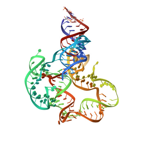
Structural insights into ligand binding and gene expression control by an adenosylcobalamin riboswitch.

Peselis, A.Serganov, A.

(2012) Nat Struct Mol Biol 19: 1182-1184

  • DOI: https://doi.org/10.1038/nsmb.2405
  • Primary Citation Related Structures: 
    4GXY

  • PubMed Abstract: 

    Coenzyme B(12) has a key role in various enzymatic reactions and controls expression of bacterial genes through riboswitches. Here we report the crystal structure of the Symbiobacterium thermophilum B(12) riboswitch bound to its ligand adenosylcobalamin. The riboswitch forms a unique junctional structure with a large ligand-binding pocket tailored for specific recognition of the adenosyl moiety and flanked by structural elements that stabilize the regulatory region and enable control of gene expression.


  • Organizational Affiliation
    • Department of Biochemistry and Molecular Pharmacology, New York University School of Medicine, New York, New York, USA.

Macromolecule Content 

  • Total Structure Weight: 61.46 kDa 
  • Atom Count: 3,685 
  • Modeled Residue Count: 163 
  • Deposited Residue Count: 172 
  • Unique nucleic acid chains: 1

Macromolecules

Find similar nucleic acids by:  (by identity cutoff) 
Entity ID: 1
MoleculeChains LengthOrganismImage
Adenosylcobalamin riboswitch172N/A
Sequence Annotations
Expand
Reference Sequence

Small Molecules

Ligands 3 Unique
IDChains Name / Formula / InChI Key2D Diagram3D Interactions
B1Z

Query on B1Z


Download:Ideal Coordinates CCD File
B [auth A],
C [auth A]
Adenosylcobalamin
C72 H101 Co N18 O17 P
NAGDYSDXWHSJMC-OUCXYWSSSA-M
IRI

Query on IRI


Download:Ideal Coordinates CCD File
D [auth A]
E [auth A]
F [auth A]
G [auth A]
H [auth A]
D [auth A],
E [auth A],
F [auth A],
G [auth A],
H [auth A],
I [auth A],
J [auth A]
IRIDIUM HEXAMMINE ION
H18 Ir N6
CGMAOQLDNKCXGK-UHFFFAOYSA-N
MG

Query on MG


Download:Ideal Coordinates CCD File
K [auth A],
L [auth A]
MAGNESIUM ION
Mg
JLVVSXFLKOJNIY-UHFFFAOYSA-N

Experimental Data & Validation

Experimental Data

  • Method: X-RAY DIFFRACTION
  • Resolution: 3.05 Å
  • R-Value Free:  0.250 (Depositor), 0.251 (DCC) 
  • R-Value Work:  0.208 (Depositor), 0.209 (DCC) 
  • R-Value Observed: 0.210 (Depositor) 
Space Group: P 31 2 1
Unit Cell:
Length ( Å )Angle ( ˚ )
a = 96.633α = 90
b = 96.633β = 90
c = 132.684γ = 120
Software Package:
Software NamePurpose
MAR345data collection
PHENIXmodel building
PHENIXrefinement
HKL-2000data reduction
HKL-2000data scaling
PHENIXphasing

Structure Validation

View Full Validation Report


Ligand Structure Quality Assessment 


Entry History 

Deposition Data

Revision History  (Full details and data files)

  • Version 1.0: 2012-10-17
    Type: Initial release
  • Version 1.1: 2012-11-21
    Changes: Database references
  • Version 1.2: 2017-11-15
    Changes: Refinement description
  • Version 1.3: 2024-02-28
    Changes: Data collection, Database references, Derived calculations